Most people have experienced the same strange certainty: around 11 PM, a wave of sleepiness arrives almost on schedule, regardless of what they were doing an hour before. And then, just as reliably, it lifts. By 1 AM, a second wind can make sleep feel impossible again. This isn't random. It reflects the output of two distinct biological systems operating in parallel — and understanding what those systems actually are, at the level of neurons and molecules, changes how you read almost everything else about sleep.
Popular accounts of circadian rhythms tend to stop at the observation that "light resets your body clock" and that "melatonin rises at night." Both statements are true, but they describe the outputs of a system whose internal architecture is far more interesting — and far more relevant to understanding why disruptions like shift work cause metabolic harm, why jet lag resolves differently than insomnia, and why some people are constitutionally unable to fall asleep before 2 AM no matter how tired they feel.
This article covers the biology directly: the anatomy of the master clock, the molecular feedback loop that runs inside it, the two-process model that explains how the clock interacts with sleep pressure, the physiological signals the clock produces, and why peripheral organs run their own clocks that can fall out of sync with the central one. The goal is mechanistic understanding, not a checklist.
The Master Clock: SCN Anatomy and Light Entrainment
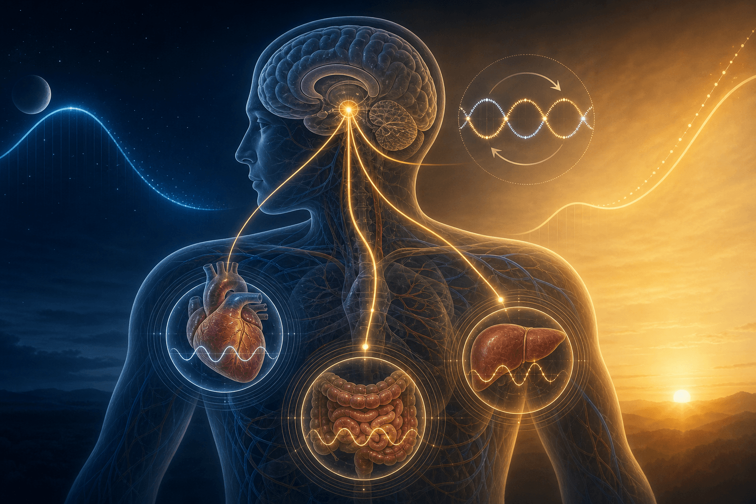
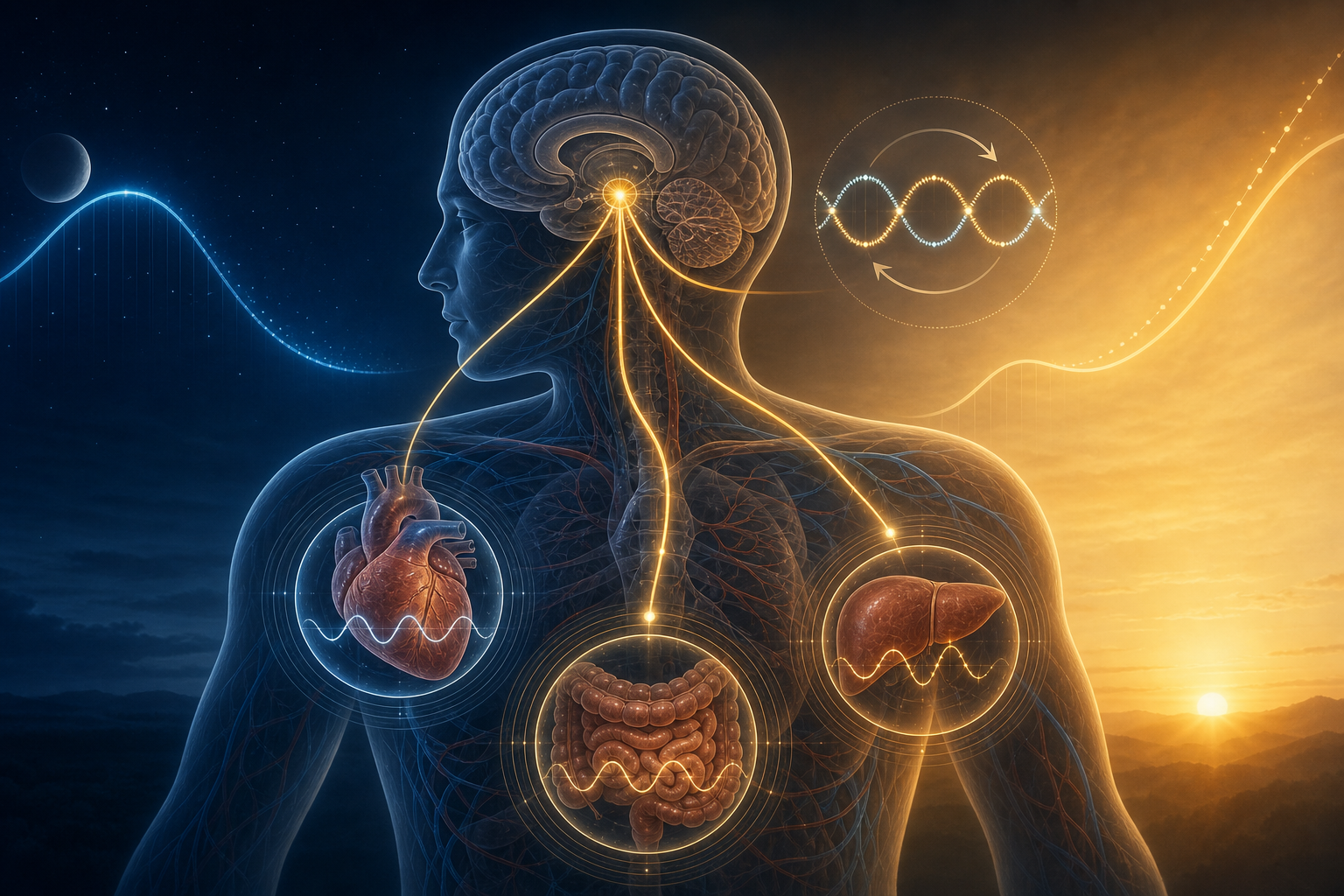
The circadian pacemaker in humans is the suprachiasmatic nucleus (SCN) — a paired structure in the hypothalamus, sitting directly above the optic chiasm. It contains roughly 20,000 neurons organized into two functionally distinct zones. The ventrolateral core, populated by neurons that express vasoactive intestinal peptide (VIP), receives direct retinal projections and is responsible for processing light signals and synchronizing the network. The dorsomedial shell, populated by neurons that express arginine vasopressin (AVP), determines the period of the ensemble rhythm — how long the SCN's oscillation actually takes to complete one cycle.
Left entirely to itself, without any external time cues, the human SCN runs on a period that averages approximately 24.18 hours — not exactly 24. This has been confirmed under strictly controlled lighting conditions. The practical consequence is that the SCN must be reset daily to stay synchronized with the solar day, and it does this primarily through light.
The light signal reaches the SCN via a dedicated pathway called the retinohypothalamic tract (RHT). The photoreceptors that drive this pathway are not the rod and cone cells used for visual perception. They are a specialized subset of retinal ganglion cells that contain their own photopigment, melanopsin, making them intrinsically photosensitive — referred to as ipRGCs (intrinsically photosensitive retinal ganglion cells). These cells are maximally sensitive to short-wavelength blue light (around 480 nm). Experimental studies have confirmed that eliminating these cells abolishes circadian photoentrainment entirely, even when rod and cone function is preserved.
The SCN receives two additional inputs beyond the RHT. The geniculohypothalamic tract carries signals from the thalamic intergeniculate leaflet, which integrates both photic and non-photic information. Serotonergic projections from the raphe nuclei provide a third input channel, modulating the SCN's responsiveness to light based on behavioral state. Together, these three pathways allow the SCN to integrate light, arousal, and activity information — though light via the RHT remains the dominant entraining signal.
Once entrained, the SCN coordinates timing across the brain and body. Its primary output pathway runs through the subparaventricular zone (SPZ) to the dorsomedial hypothalamic nucleus (DMH), which relays timing information to regulate sleep, locomotor activity, body temperature, and reproductive functions. A separate output pathway through the paraventricular nucleus (PVN) controls the hormonal outputs described later in this article.
The Molecular Clockwork: How CLOCK, BMAL1, PER, and CRY Build a 24-Hour Timer

Before introducing the specific molecules, it helps to have an analogy. Imagine a thermostat that controls its own furnace, but with a twist: when the furnace heats the room, it also sets a timer that will eventually shut the furnace off. Once the room cools below a threshold, the furnace restarts. The cycle takes a fixed amount of time to complete — not because of an external clock, but because the heating and cooling dynamics are built into the system itself. That is roughly what happens inside SCN neurons at the molecular level.
The core mechanism is called a transcription-translation feedback loop (TTFL). Here is how it works in plain terms, followed by the gene names:
- Two proteins — CLOCK (think of it as the "activator") and BMAL1 (its partner, also an activator) — join together to form a complex. This CLOCK/BMAL1 heterodimer binds to specific DNA sequences called E-boxes and switches on the transcription of two families of genes: the Period genes (PER1, PER2, PER3) and the Cryptochrome genes (CRY1, CRY2).
- Over the course of several hours, PER and CRY proteins accumulate in the cytoplasm of the cell. They then form their own complex and move into the nucleus — the cell's control center.
- Once inside the nucleus, the PER/CRY complex binds to CLOCK and BMAL1, blocking their ability to activate gene transcription. This shuts off production of new PER and CRY proteins — the furnace turns off.
- Existing PER and CRY proteins are gradually degraded by cellular enzymes over several more hours. As their concentrations fall, the inhibition on CLOCK/BMAL1 lifts, and the cycle begins again.
The total duration of one cycle — from CLOCK/BMAL1 activation through PER/CRY accumulation, nuclear entry, inhibition, and degradation — is approximately 24 hours. The precision of this timing depends on the rates of protein synthesis, phosphorylation (chemical modification that affects stability), and degradation. Mutations that alter these rates shift the period of the clock, which is exactly what happens in familial advanced sleep phase syndrome, where a mutation in a kinase that phosphorylates PER proteins causes them to degrade faster, shortening the cycle and pushing sleep onset abnormally early.
A secondary feedback loop adds robustness to this system. CLOCK/BMAL1 also drives the transcription of genes called REV-ERB (a repressor — it suppresses gene expression) and ROR (an activator — it promotes gene expression). Both REV-ERB and ROR compete to regulate the transcription of BMAL1 itself. When REV-ERB levels are high, BMAL1 transcription is suppressed; when ROR dominates, BMAL1 transcription rises. This secondary loop does not generate the oscillation on its own, but it stabilizes the amplitude and period of the primary loop, making the clock more resistant to noise and environmental perturbations.
One further implication of this molecular architecture: because the TTFL runs inside virtually every cell in the body — not just SCN neurons — nearly half of all protein-coding genes show circadian-dependent expression patterns in at least one tissue. The SCN is the master coordinator, but the molecular clock is a cellular property, not a brain-exclusive one. This becomes critically important when we discuss peripheral clocks.
The Two-Process Model: How the Clock and Sleep Pressure Interact

Knowing that the SCN runs a molecular clock is necessary but not sufficient to explain why you fall asleep when you do. The SCN's circadian signal is only one of two systems governing sleep propensity. The other is the homeostatic sleep drive — and the relationship between them was formalized in 1982 by Alexander Borbély as the two-process model, which remains the foundational framework in sleep science.
Process S: Homeostatic Sleep Pressure
Process S represents the accumulation of sleep need during wakefulness. Its most studied molecular correlate is adenosine — a byproduct of neuronal energy metabolism that builds up in the extracellular space of the brain during sustained wakefulness. As adenosine concentrations rise, it binds to adenosine A1 receptors in wake-promoting brain regions, progressively suppressing their activity. The longer you stay awake, the higher the adenosine load, and the more intense the pressure to sleep becomes.
Process S can be measured empirically using EEG. During non-REM sleep, the electroencephalogram shows slow, high-amplitude oscillations called slow-wave activity (SWA, typically 0.5–4 Hz). SWA intensity at the beginning of a sleep period reflects how much homeostatic pressure had accumulated before sleep onset — it is higher after sleep deprivation and lower after a nap. As sleep continues, SWA declines exponentially, tracking the dissipation of adenosine and the recovery of homeostatic balance.
Caffeine works precisely at this level: it is a competitive antagonist at adenosine receptors, blocking adenosine's ability to signal sleep pressure without actually reducing adenosine concentrations. This is why caffeine can delay sleepiness but tends to produce a rebound in fatigue once it wears off — the adenosine that was blocked is still present and resumes signaling.
Process C: The Circadian Alerting Signal
Process C represents the circadian pacemaker's output as it relates to sleep and wakefulness. Critically, the circadian signal does not simply track with or amplify sleep pressure — it actively opposes it during the biological day. In the late afternoon and early evening, the SCN generates a strong alerting signal that counteracts the rising adenosine load, which is why people often feel most alert around 6–8 PM despite having been awake for 12 or more hours. This is sometimes called the "wake maintenance zone" — a circadian-driven window of high alertness that temporarily overrides homeostatic pressure.
As the evening progresses and the SCN transitions toward its night-oriented state, the alerting signal falls away. With both high adenosine pressure and a declining circadian alerting signal converging, sleep onset becomes almost inevitable — which is the experience of that predictable evening sleepiness described at the start of this article.
How S and C Interact
The two processes are not independent. A landmark forced desynchrony study by Dijk and Czeisler in 1995 — in which participants were placed on artificial 28-hour "days" that progressively shifted their sleep timing relative to their circadian phase — confirmed that homeostatic and circadian factors contribute quasi-equally to most sleep variables, including sleep onset latency, sleep duration, and the distribution of slow-wave sleep across the night.
There is also a feedback relationship between the two systems. Elevated sleep pressure (high adenosine) can dampen the SCN's responsiveness to light. The proposed mechanism involves adenosine inhibiting glutamate release from the retinohypothalamic tract at the SCN synapse, reducing the magnitude of light-induced phase shifts. This means that severe sleep deprivation — by raising adenosine — can partially blunt the clock's ability to reset itself, creating a compounding disruption rather than two independent problems.
Key Circadian Output Signals: Melatonin, Cortisol, and Core Body Temperature
The SCN communicates its timing information to the rest of the body through several output signals. Three of them — melatonin, cortisol, and core body temperature — are particularly well-characterized and illustrate how the clock translates molecular oscillation into whole-body physiology.
Melatonin: A Darkness Signal, Not a Sleep Drug
Melatonin is produced and secreted by the pineal gland, a small structure deep in the brain. The anatomical pathway controlling its secretion runs from the SCN to the paraventricular nucleus (PVN) of the hypothalamus, then down through the spinal cord to the intermediolateral nucleus, then to the superior cervical ganglion, and finally via sympathetic nerve fibers to the pineal gland. When the SCN is active during the day, it sends inhibitory (GABAergic) signals through this pathway that suppress pineal melatonin production. As SCN activity falls at night, this inhibition is released, and the superior cervical ganglion activates the pineal gland to secrete melatonin.
This pathway explains why light at night suppresses melatonin: light activates ipRGCs, which activate the SCN, which reactivates the inhibitory signal to the pineal gland. The suppression is not a side effect — it is the mechanism. The pineal gland is essentially reading the SCN's activity state and broadcasting that information as a hormonal darkness signal.
The Cortisol Awakening Response
Cortisol follows a sharply timed circadian rhythm driven by the SCN through the hypothalamic-pituitary-adrenal (HPA) axis. Levels are at their lowest in the first half of the night and begin rising in the hours before habitual wake time. The most striking feature of this pattern is the cortisol awakening response (CAR): a sharp, rapid increase in cortisol concentrations occurring approximately 30 to 45 minutes after waking. This rise is driven by SCN → PVN → HPA axis signaling and appears to serve a preparatory function — mobilizing glucose, activating immune function, and priming cognitive systems for daytime activity. The CAR is distinct from the general circadian cortisol rhythm and is blunted by sleep deprivation and chronic stress, which has made it a useful biomarker in psychoneuroendocrinology research.
Core Body Temperature
Core body temperature follows a robust circadian rhythm driven by the SCN via the sub-paraventricular zone and the medial preoptic area, which controls heat dissipation through peripheral vasodilation. Temperature begins falling in the evening, reaches a nadir of approximately 1°C below daytime values around 4 to 5 AM, and then rises toward wake time. The fall in core temperature is not merely a consequence of sleep — it is mechanistically necessary for sleep initiation. The medial preoptic area, which is the primary sleep-promoting region of the brain, is activated by the falling temperature signal. Experimental studies that artificially warm the skin (promoting heat dissipation and thus cooling the core) reliably accelerate sleep onset and increase slow-wave sleep.
| Output Signal | Source | Pathway | Circadian Pattern | Functional Role |
|---|---|---|---|---|
| Melatonin | Pineal gland | SCN → PVN → intermediolateral nucleus → superior cervical ganglion → pineal | Rises at dusk, peaks mid-night, falls before dawn | Darkness signal and circadian phase marker; suppressed by light |
| Cortisol | Adrenal cortex | SCN → PVN → HPA axis (CRH → ACTH → cortisol) | Lowest mid-night; sharp CAR 30–45 min post-wake; falls through afternoon | Metabolic and immune preparation for wakefulness; CAR primes cognition |
| Core body temperature | Peripheral vasodilation (heat dissipation) | SCN → sub-paraventricular zone → medial preoptic area → autonomic | Falls ~1°C through evening; nadir ~4–5 AM; rises toward wake | Mechanistically necessary for sleep initiation; activates sleep-promoting neurons |
Peripheral Clocks and Internal Misalignment
The SCN is the master clock, but it is not the only clock. The same CLOCK/BMAL1/PER/CRY transcription-translation feedback loop that runs in SCN neurons also runs in cells throughout the body — in the liver, heart, gut, pancreas, kidneys, skin, adrenal gland, and circulating immune cells (peripheral blood mononuclear cells, or PBMCs). These peripheral oscillators are semi-autonomous: they can sustain rhythmic gene expression on their own for several cycles even when isolated from the SCN.
The critical difference between the SCN clock and peripheral clocks lies in what entrains them. The SCN is predominantly entrained by light via the retinohypothalamic tract. Peripheral clocks, by contrast, are predominantly entrained by feeding and fasting cycles. The liver clock, for example, tracks when food arrives and adjusts its metabolic gene expression accordingly — timing the production of digestive enzymes, gluconeogenic enzymes, and lipid metabolism regulators to anticipate when nutrients will be available.
Under normal conditions, when meals are consumed during the active phase (daytime for humans), the feeding-entrained peripheral clocks and the light-entrained SCN are synchronized. The system operates in internal alignment: the liver expects glucose when the brain expects activity, and both are timed to the same solar day.
When meals are shifted to the biological night — as happens during night shift work, or with habitual late eating — peripheral clocks reorient toward the new feeding schedule while the SCN remains anchored to the light-dark cycle. This creates internal circadian misalignment: the central clock and the peripheral clocks are running on different schedules within the same body.
The practical consequence of this biology is that circadian disruption is not simply a matter of feeling tired at the wrong time. The metabolic machinery of the liver, pancreas, and gastrointestinal tract is timed to expect nutrients, process glucose, and regulate insulin sensitivity during the active phase. When food arrives during the biological night, this machinery is out of phase with the nutritional load. Over time, this mismatch is associated with increased risk of metabolic syndrome, impaired glucose tolerance, and type 2 diabetes — not because of poor food choices, but because of mistimed ones.
- Peripheral clocks in the liver, gut, pancreas, heart, and skin run the same CLOCK/BMAL1/PER/CRY loop as the SCN.
- Peripheral clocks are primarily entrained by feeding timing, not by light.
- Mistimed meals (eating during the biological night) can shift peripheral clocks away from the SCN, creating internal misalignment.
- Internal misalignment — not just external misalignment with the light-dark cycle — is the proposed mechanism linking shift work to metabolic disease.
- Without specific interventions, only approximately 25% of night workers show full circadian adaptation of their central clock to a night-oriented schedule.
Clinical Significance: Why Insomnia and Circadian Rhythm Disorders Are Not the Same Condition
One of the most consequential misunderstandings in popular sleep content is the conflation of insomnia with circadian rhythm sleep disorders. They are not variants of the same problem. They arise from different mechanisms, present with different patterns, and respond to different treatments. The biology covered in this article makes the distinction clear.
Insomnia — specifically chronic insomnia disorder — is primarily characterized by hyperarousal: an elevated physiological and cognitive activation state that interferes with the ability to initiate or maintain sleep. The circadian clock (Process C) is typically functioning normally in people with insomnia; the problem lies in dysregulated sleep homeostasis and arousal systems. The clock is in the right place; the system that should be winding down for sleep is not cooperating.
Circadian rhythm sleep disorders (CRSDs) involve a different problem entirely: the clock itself is shifted or misaligned relative to the desired or socially required sleep schedule. In delayed sleep-wake phase disorder (DSPD), the circadian pacemaker is shifted later — melatonin onset, core temperature nadir, and the cortisol awakening response all occur several hours later than in the general population. The person is not hyperaroused; they are simply trying to sleep at a time when their biology is in the circadian "wake maintenance zone." When allowed to sleep on their own schedule — sleeping at 3 AM and waking at 11 AM, for example — they often sleep normally in terms of duration and architecture.
Shift work disorder shares the circadian misalignment mechanism but is imposed externally: work schedules require activity during the biological night, and the SCN does not adapt quickly enough (or, for many workers, at all). Non-24-hour sleep-wake rhythm disorder, most prevalent in blind individuals who lack photic entrainment, represents a failure of the entrainment mechanism itself — the clock free-runs at its intrinsic ~24.18-hour period with no daily reset, causing sleep timing to drift progressively around the clock.
| Condition | Primary Mechanism | Process Affected | Sleep Timing Pattern | Distinguishing Feature |
|---|---|---|---|---|
| Chronic insomnia disorder | Hyperarousal; dysregulated sleep homeostasis | Process S and arousal systems | Difficulty initiating or maintaining sleep at a normal or desired time | Clock phase is typically normal; the problem is arousal, not timing |
| Delayed sleep-wake phase disorder (DSPD) | Shifted circadian pacemaker (delayed) | Process C | Inability to fall asleep until very late; difficulty waking at conventional times | Normal sleep architecture and duration when allowed to sleep on own schedule |
| Shift work disorder | External schedule forces activity during biological night; partial or absent clock adaptation | Process C and internal alignment | Insomnia during required sleep periods; excessive sleepiness during required wake periods | Circadian phase remains day-oriented despite night work schedule |
| Non-24-hour sleep-wake disorder | Loss of photic entrainment; clock free-runs | Process C entrainment | Sleep timing drifts progressively later (or earlier) over days to weeks | Most common in totally blind individuals; clock period is intact but unentrainable |
Beyond sleep quality, the health consequences of chronic circadian misalignment extend considerably further. The International Agency for Research on Cancer (IARC) classifies night shift work as probably carcinogenic to humans, based on evidence linking long-term night shift work to elevated risk of breast and other cancers. Night shift work is also associated with increased risk of metabolic syndrome, type 2 diabetes, and cardiovascular disease — consistent with the peripheral clock misalignment mechanisms described in the preceding section.
The clinical architecture of circadian rhythm disorders — definitions, diagnostic criteria, and treatment protocols — is covered in detail in the Sleep Conditions section of this site. The mechanistic grounding provided here is intended to make that clinical content more interpretable, not to substitute for it.
Key Takeaways
- The suprachiasmatic nucleus (SCN) is a paired hypothalamic structure of approximately 20,000 neurons that acts as the master circadian pacemaker. Its endogenous period averages ~24.18 hours and requires daily photic entrainment via melanopsin-expressing ipRGCs along the retinohypothalamic tract.
- At the molecular level, the circadian clock operates through a transcription-translation feedback loop: CLOCK and BMAL1 proteins drive transcription of PER and CRY genes; the resulting PER/CRY proteins inhibit CLOCK/BMAL1 activity, completing a self-sustaining ~24-hour cycle. A secondary REV-ERB/ROR loop stabilizes the oscillation.
- Sleep timing and depth are determined by the interaction of two processes: Process S (homeostatic sleep pressure, built by adenosine during wakefulness and reflected in EEG slow-wave activity) and Process C (the circadian pacemaker's alerting signal, which opposes sleepiness during the day and withdraws at night). Neither process alone determines sleep — their interaction does.
- Melatonin is a darkness signal and circadian phase marker, not a sleep-inducing hormone. It is secreted by the pineal gland when SCN inhibition falls at night, via the SCN → PVN → intermediolateral nucleus → superior cervical ganglion pathway. Dim light melatonin onset (DLMO) is the clinical gold standard for measuring circadian phase.
- The cortisol awakening response (a sharp cortisol rise 30–45 minutes post-wake, driven by SCN → PVN → HPA axis signaling) and the nightly ~1°C drop in core body temperature (driven by SCN → sub-paraventricular zone → medial preoptic area) are two additional circadian output signals with direct functional roles in preparing the body for wakefulness and sleep initiation, respectively.
- Most cells and organs contain autonomous circadian oscillators using the same CLOCK/BMAL1/PER/CRY machinery as the SCN. Peripheral clocks are preferentially entrained by feeding timing rather than light. Mistimed meals can decouple peripheral clocks from the SCN, creating internal circadian misalignment — the proposed mechanism linking shift work and late eating to metabolic disease.
- Insomnia and circadian rhythm sleep disorders are mechanistically distinct conditions. Insomnia involves hyperarousal and dysregulated sleep homeostasis (Process S); circadian rhythm sleep disorders such as DSPD and shift work disorder involve a shifted or misaligned circadian pacemaker (Process C). This distinction has direct implications for which interventions are appropriate.
Comments
Join the discussion with an anonymous comment.