
Mechanism explainer
How Frontotemporal Dementia Disrupts Sleep
Explains why frontotemporal dementia produces a distinct sleep pattern: increased total sleep time but poor efficiency, high fragmentation, and morning inactivity — linked to frontal lobe atrophy rather than the circadian rhythm changes seen in other dementias.
In behavioral variant frontotemporal dementia, the sleep problem often begins with a contradiction: the person may spend far more time in bed and still leave the household feeling as if nobody slept. That is not just a caregiver impression. In actigraphy data from people with behavioral variant frontotemporal dementia, longer sleep sat beside poorer sleep efficiency, more time awake after sleep began, more movement at night, and a notable drop in morning activity.[1]
That pattern is the clearest answer to how frontotemporal dementia disrupts sleep. In bvFTD, the trouble is not simply that the sleep schedule shifts later or that evenings become agitated. The disease appears to interfere with frontal brain systems that help keep sleep and wakefulness organized. The result can look deceptively like “sleeping all the time,” when the closer measurement is: more time in bed, more minutes asleep, but a weaker boundary between sleep and wake.

The paradox: longer nights, less efficient sleep
A 2024 actigraphy study gives this paradox unusually concrete shape. Researchers compared 27 people with behavioral variant frontotemporal dementia with 25 controls. The bvFTD group spent about 566 minutes in bed, compared with about 444 minutes in controls. They also slept longer in total: about 463 minutes versus about 377 minutes.[1]
If those were the only numbers, a family might hear reassurance: more sleep should mean more recovery. But the same study showed the opposite side of the night. Sleep efficiency was lower in bvFTD, about 81.9% compared with 88.7% in controls, and wake after sleep onset was much higher: about 69 minutes versus 29 minutes.[1] In ordinary household terms, the person was in bed much longer, slept more in total, and still spent far more of the night awake after sleep had supposedly started.
| Actigraphy measure | bvFTD group | Control group | What the measure helps explain |
|---|---|---|---|
| Time in bed | About 566 minutes | About 444 minutes | Why caregivers may see the person as sleeping or resting for much of the night |
| Total sleep time | About 463 minutes | About 377 minutes | Why “more sleep” can be a real measurement, not just an impression |
| Sleep efficiency | About 81.9% | About 88.7% | Why longer sleep does not necessarily mean consolidated, restorative sleep |
| Wake after sleep onset | About 69 minutes | About 29 minutes | Why the night can feel broken even when total sleep time is high |
The study also found that 85.2% of the bvFTD group met Pittsburgh Sleep Quality Index criteria for poor sleep quality, compared with 48% of controls.[1] That survey result matters, but the movement data are especially useful because they show where the night breaks apart. A person can accumulate many minutes of sleep across a long stretch in bed, yet keep surfacing into wakefulness, moving, or failing to settle into a stable sleep period.
This is one reason broad phrases such as “disturbed sleep” can feel so inadequate. The caregiver is not just reporting a vague disturbance. They may be seeing a person who goes to bed early, stays in bed late, wakes repeatedly, moves during the night, and then starts the morning with unusually low activity. Those details point toward a specific sleep phenotype rather than a generic dementia sleep complaint.
The morning can be part of the sleep problem
The same actigraphy work found a distinctive daily activity pattern: lower activity in the morning window from 6:30 to 11:30 AM, and higher activity at night from 2:00 to 5:20 AM.[1] That combination is easy to misread. A flat morning after a long night in bed can look like apathy alone, depression alone, medication sedation, or refusal to get going. Any of those may still need consideration, but the sleep data show that the day may be starting from a fragmented night rather than from true rest.
For a partner or adult child, the practical burden sits in the timing. Nighttime activity demands surveillance when the caregiver is least able to think clearly. Morning inactivity then arrives when the household needs bathing, meals, appointments, or supervision to restart. The person with bvFTD is not simply choosing an inconvenient schedule. Their sleep-wake organization may be losing the structure that normally separates nighttime rest from daytime activation.
Why frontal lobe atrophy belongs in the sleep conversation
Frontotemporal dementia is named for the frontal and temporal brain regions it damages, but sleep discussions often treat that damage as background scenery. In bvFTD, the frontal findings deserve to be placed closer to the center. Filardi and colleagues reported that increased sleep duration correlated with reduced cortical thickness in the superior frontal gyrus, orbitofrontal cortex, and anterior cingulate gyrus.[1]
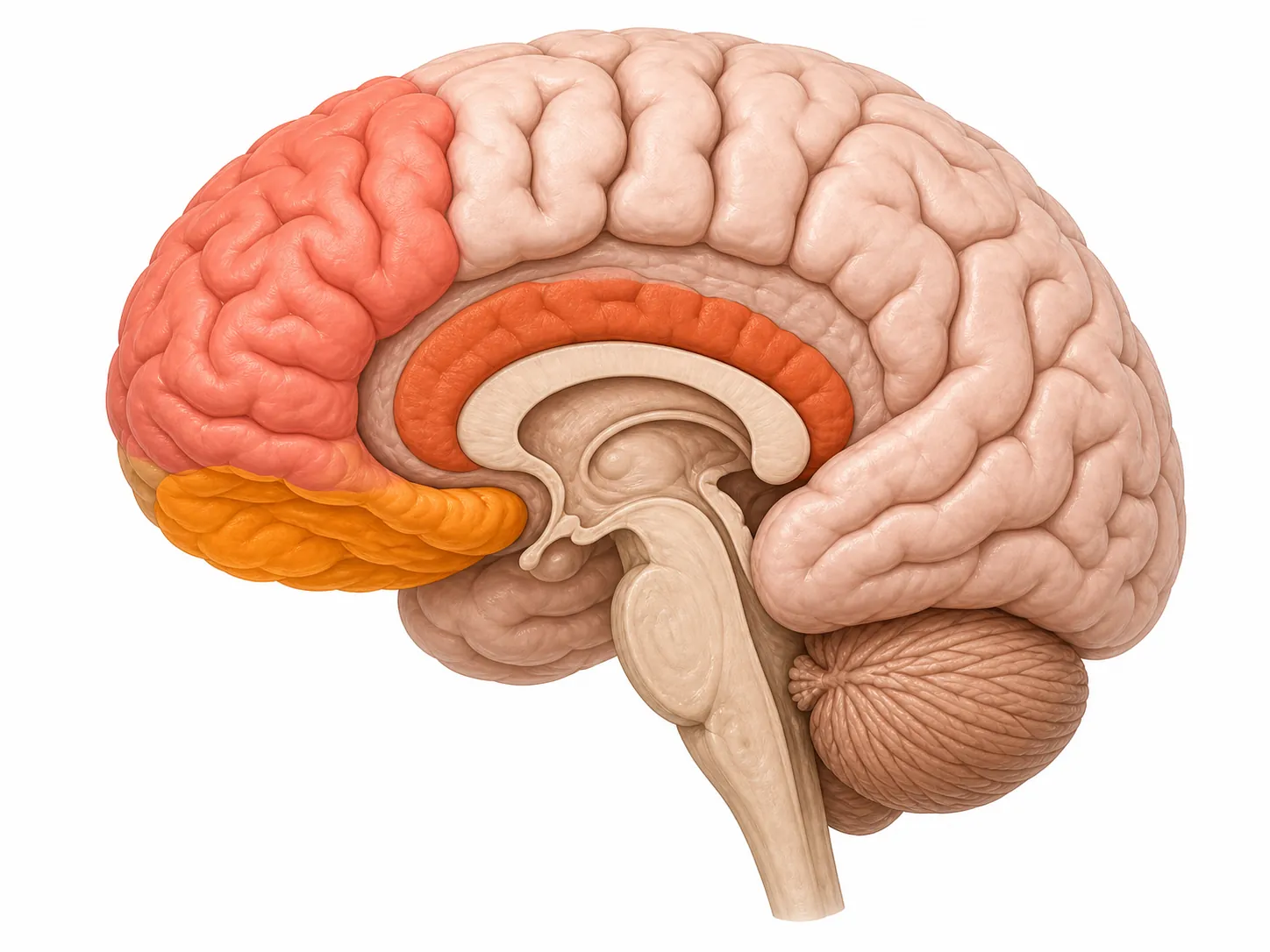
Those regions are not sleep switches in the simple sense. They are better understood as parts of a frontal control network that helps regulate behavior, attention, inhibition, and the organized transitions that make wakefulness and sleep feel like distinct states. When these areas thin, a person may not merely become disinhibited or apathetic during the day. The continuity of the sleep period itself may also become harder to maintain.
The orbitofrontal cortex is often discussed in relation to impulse control and behavioral regulation. The anterior cingulate is involved in motivation, monitoring, and the allocation of effort. The superior frontal region is part of broader prefrontal systems that help organize behavior over time. In sleep terms, damage across these regions offers a plausible explanation for the pattern actigraphy records: extended time in bed without the normal efficiency of consolidated sleep.
The caution is important. The MRI correlation analyses in the 2024 study were exploratory and were not corrected for multiple comparisons.[1] That means the findings should not be treated as final proof that thinning in one named region directly causes one sleep metric. But they do support a more specific reading than “dementia disrupts sleep.” In bvFTD, the sleep disturbance appears tied to frontal network degeneration in a way that matches the disease’s anatomy.
There may also be a deeper sleep-wake regulation layer involving hypothalamic systems and hypocretin, also called orexin, which helps stabilize wakefulness. That possibility fits the broader biology of sleep-wake control, but it should stay in the supporting role here. The best caregiver-facing evidence for this specific bvFTD sleep pattern still comes from measured rest-activity data and the frontal cortical associations.
Why this is not just Alzheimer’s sleep disruption under another name
Comparing FTD with Alzheimer’s disease is useful only if it sharpens the distinction. Both conditions can disrupt sleep, and families living with either disease may face nights that are exhausting. But the available studies suggest that bvFTD can produce sleep disruption early and sometimes severely, without simply copying the Alzheimer’s circadian pattern.
In a 2014 Sleep Medicine study comparing 12 people with bvFTD, 17 with Alzheimer’s disease, and 20 controls, the authors concluded that sleep in FTD was “equally or even more affected by neurodegeneration” than sleep in Alzheimer’s disease, and over a “much shorter time span.”[2] The sample was small, so it should not be stretched into a universal rule. Still, it matches the clinical problem many families notice: sleep can become strange and difficult before the person appears globally late-stage.
A separate comparison of men with frontotemporal degeneration and Alzheimer disease found that the FTD group had a more fragmented rest-activity rhythm and a distinctive morning hypoactivity that was not seen in the Alzheimer group. The Alzheimer group showed a phase-delayed temperature rhythm that was absent in FTD.[3] That distinction matters because it pushes the explanation away from a one-size-fits-all dementia circadian story.
In Alzheimer’s disease, caregivers are often told to watch for circadian drift, day-night reversal, and sundowning. Those can be real and serious problems. But in bvFTD, the more revealing pattern may be the uneven distribution of activity: too much movement deep in the night, too little activation in the morning, and a long interval in bed that fails to become a clean block of restorative sleep.
A brief note on REM sleep behavior disorder
REM sleep behavior disorder is worth mentioning because it often appears in dementia sleep discussions. It involves dream enactment behaviors during REM sleep and is strongly associated with synuclein-related disorders. In FTD, however, REM sleep behavior disorder appears to be rare compared with synucleinopathies and is not the central sleep clue.[4] If vivid dream enactment, punching, kicking, or falling out of bed is prominent, that history deserves clinical attention, but it does not explain the main bvFTD pattern of long, inefficient, fragmented sleep.
When the person’s report and the night’s movement disagree
The gap between perceived sleep and measured sleep can be large enough to change the whole interpretation. A 2023 case report described a person with FTD who reported sleeping only 2.5 hours, while actigraphy showed 8 hours and 37 minutes of sleep, a discrepancy of more than 6 hours.[5] That single case does not prove that every person with FTD misperceives sleep in the same way. It does show why relying only on the patient’s estimate, or only on a caregiver’s exhausted memory of the night, can mislead everyone involved.
Objective measurement does not have to mean that every family immediately needs a device. A simple sleep diary can still help, especially if it records bedtimes, out-of-bed times, nighttime wandering or movement, naps, morning inactivity, medication changes, alcohol use, and breathing symptoms. But when the story is contradictory — “he sleeps constantly” and “he is awake all night” — actigraphy can make the contradiction legible instead of forcing the family to choose which impression is true.
The measurement also protects the person with dementia. Fragmented nights can look like stubbornness, laziness, or deliberate noncooperation from the outside. A rest-activity record reframes the problem as a pattern the brain is producing, not a moral failure the person is choosing.
Do not let the FTD explanation hide treatable sleep disorders
A frontal-network explanation should not become a wastebasket explanation. The 2024 bvFTD study also found higher STOP-Bang scores, a screening measure for obstructive sleep apnea risk, in the bvFTD group.[1] That does not mean every person with FTD has sleep apnea, and STOP-Bang is not the same as a diagnostic sleep study. It does mean that breathing-related sleep disruption should not be waved away as “just the dementia.”
This boundary is practical. Loud snoring, witnessed pauses in breathing, gasping, morning headaches, marked daytime sleepiness, or worsening nighttime confusion may justify asking a clinician whether sleep apnea evaluation is appropriate. Restless legs, pain, urinary frequency, medication effects, alcohol, depression, and environmental disruption can also coexist with bvFTD. The presence of a disease-specific sleep phenotype does not make ordinary sleep medicine irrelevant.
Large population data add another reason to take very long sleep seriously without overinterpreting it. A 2025 UK Biobank research snapshot described data from 396,918 participants in which extremely long sleep duration, defined as more than 9 hours, was associated with lower total brain volume, impaired executive function, and links to FTD/ALS-FTSD diagnosis.[6] That kind of association cannot tell a family that long sleep caused the disease or that one sleeper is developing FTD. It does support the narrower point: major changes in sleep duration and executive function belong in the clinical conversation.
What the pattern means in bvFTD
The strongest evidence applies to behavioral variant frontotemporal dementia, not every FTD variant. Primary progressive aphasia and other forms of FTD are less studied for sleep, and it would be premature to assume that all variants follow the same measured pattern. Even in bvFTD, the main studies are modest in size. Their consistency is useful; their limits are real.
Within those limits, the calibrated judgment is clear enough to help families interpret what they are seeing. In bvFTD, disrupted sleep is best understood as a frontal-network sleep-continuity problem. Atrophy involving regions such as the superior frontal gyrus, orbitofrontal cortex, and anterior cingulate is associated with longer sleep duration, while actigraphy shows the lived consequence: more time in bed, lower sleep efficiency, more wake after sleep onset, more nighttime activity, and quieter mornings.
So when someone with bvFTD seems to sleep all the time and still leaves the household unrested, the contradiction may be the clue. The night is longer, but less continuous. The morning arrives, but the body is not fully activated. The caregiver’s observation is not too vague to matter; it is often the first rough version of a pattern that actigraphy can measure.
References
- Sleep and circadian rhythm disruptions in behavioral variant frontotemporal dementia, Alzheimer’s & Dementia, 2024.
- Sleep in frontotemporal dementia is equally or possibly more disrupted, and at an earlier stage, when compared to sleep in Alzheimer's disease, Sleep Medicine, 2014.
- Differential Circadian Rhythm Disturbances in Men with Alzheimer Disease and Frontotemporal Degeneration, JAMA Psychiatry, 2001.
- Sleep Disturbances in Frontotemporal Dementia, Current Neurology and Neuroscience Reports, 2016.
- Sleep State Misperception in Frontotemporal Dementia, Primary Care Companion for CNS Disorders, 2023.
- Research snapshot: Could sleep changes signal the early stages of MND or FTD?, UK MND Research Institute, 2025.
Supports these guides
Spot an error or have clinical feedback?
Because this article covers clinical, medication, or safety information, we use a moderated correction channel instead of open public comments. Let us know if something about “How Frontotemporal Dementia Disrupts Sleep” needs a closer look.
Send feedback on this article