
Why 'Just Tired' Is a Profound Understatement
When people describe what poor sleep does to them, they tend to reach for words like "foggy" or "drained" — language that implies a general dimming of mental capacity, as if every part of the brain turned down equally. Neuroimaging evidence tells a different story.
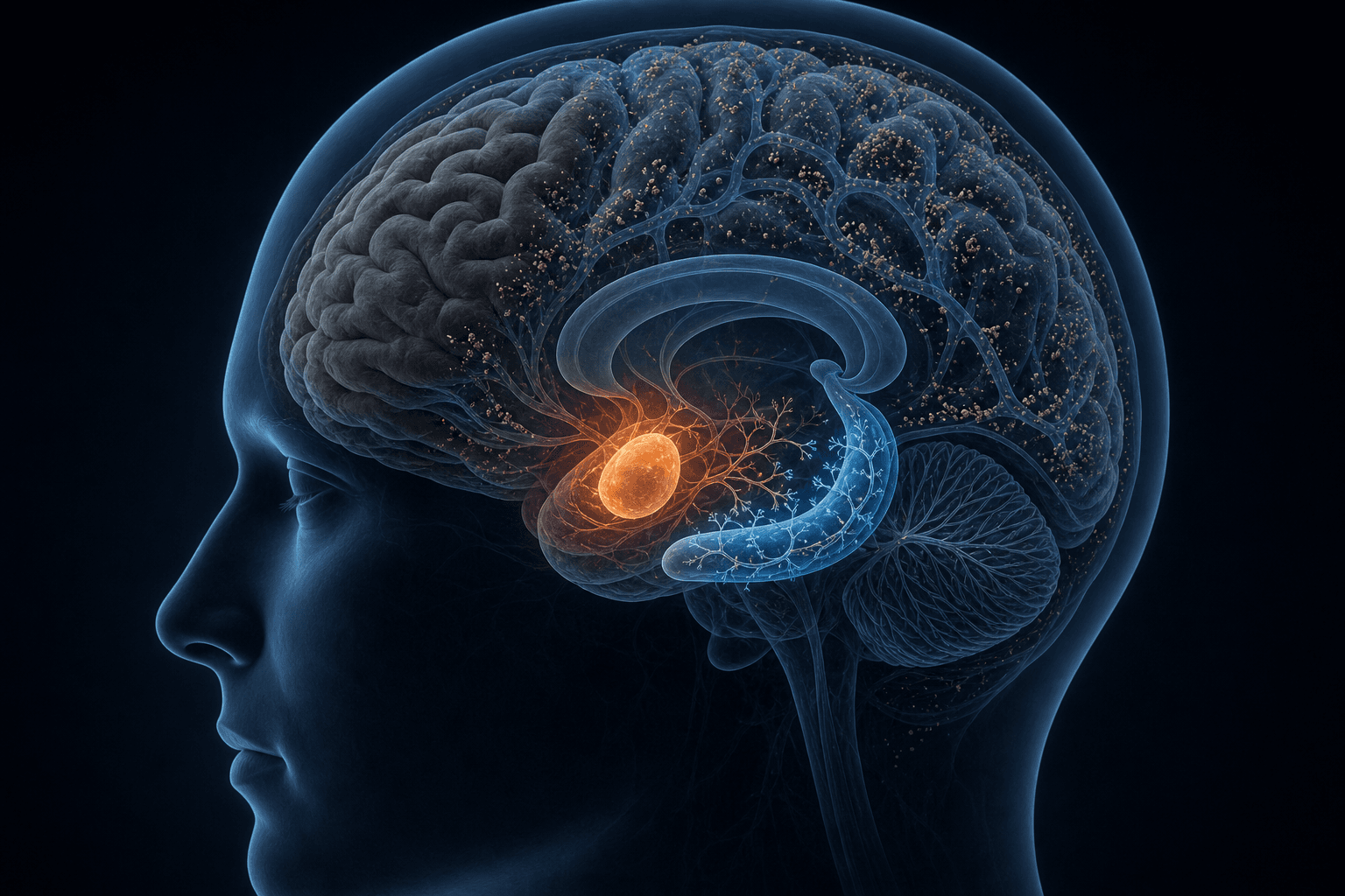
fMRI and PET studies of sleep-deprived brains do not show uniform suppression. They show selective regional dysfunction: some areas go offline while others become pathologically overactive. The prefrontal cortex and hippocampus are disproportionately impaired. The amygdala, by contrast, becomes hyperreactive — its activity amplified, its regulatory connections weakened. Meanwhile, the brain's default mode network begins intruding on tasks it should be disengaged from, producing an erratic, unpredictable pattern of attentional collapse.
This regional specificity is why sleep deprivation is not simply a matter of being slower. Different cognitive failures — forgetting something within minutes of reading it, snapping at a minor irritation, losing focus mid-sentence even in a quiet room — have distinct neurobiological explanations. Each one maps onto a specific mechanism, in a specific brain region, with a specific molecular basis.
The Prefrontal Cortex: The First Region to Fail
The prefrontal cortex (PFC) — particularly the dorsolateral prefrontal cortex (DLPFC) — governs the cognitive functions most people associate with clear thinking: planning, working memory, impulse control, moral judgment, and the ability to assess risk rather than just react to it. It is also the first major region to show measurable impairment under sleep deprivation.
fMRI studies show reduced DLPFC and intraparietal sulcus activation during attention and working-memory tasks after sleep deprivation. PET imaging reveals relative regional glucose decreases concentrated in the prefrontal and posterior parietal cortex — the brain is consuming less metabolic fuel in the regions responsible for executive control. Functional near-infrared spectroscopy (fNIRS) and transcranial Doppler (TCD) studies have documented impaired cortical hemodynamic responses and reduced cerebral blood flow in the prefrontal cortex during cognitive tasks following 24 hours of wakefulness, providing a vascular mechanism that helps explain the fMRI signal changes.
A second mechanism operates at the neurochemical level. As wakefulness extends, adenosine accumulates across the brain — this is the "sleep pressure" signal that normally builds across the day. In the striatum, elevated adenosine interferes with dopamine D2 and D3 receptor signaling. Because dopamine in the striatum is central to reward-based decision-making and the evaluation of risk vs. gain, this disruption compounds the prefrontal impairment: not only is the evaluative cortex running with reduced activity, but the reward-processing circuitry driving decisions is simultaneously dysregulated toward short-term gain and away from considered risk assessment.
The practical consequence is that executive function deteriorates in a predictable sequence. Planning becomes effortful and shallow. Working memory — the mental workspace that holds information while you use it — degrades. Impulse control weakens. And moral judgment, which depends on the integration of rule-based reasoning with emotional context that the PFC coordinates, becomes slower and less reliable. These are not subjective complaints. They are measurable neural events.
- Reduced DLPFC activation during attention and working-memory tasks (fMRI evidence)
- Prefrontal and posterior parietal glucose decreases (PET evidence)
- Impaired neurovascular coupling and reduced cerebral blood flow in the prefrontal cortex (fNIRS and TCD evidence)
- Dopamine D2/D3 receptor downregulation in the striatum via adenosine accumulation, impairing reward-based risk assessment
The Hippocampus: Why You Cannot Form New Memories During Deprivation
There is a common assumption that sleep deprivation impairs memory primarily at retrieval — that information enters the brain normally, but cannot be found later. This is incorrect, or at least incomplete. Sleep deprivation blocks new memory encoding during the deprivation period itself. Information that does not encode properly cannot be retrieved, because it was never durably stored to begin with.
The mechanism begins at the synapse. Hippocampal long-term potentiation (LTP) — the cellular process by which synaptic connections are strengthened to encode new memories — requires NMDA receptor activation and the calcium influx it triggers. Under sleep deprivation, the surface expression of NR2A and NR1 subunits of NMDA receptors is reduced, and the NMDA-to-AMPA receptor ratio at hippocampal synapses decreases. The result is attenuated calcium influx and a measurable impairment of LTP induction.
This initiates a cascade of downstream failures. Phosphodiesterase 4 (PDE4) activity — specifically the PDE4A5 isoform — increases during sleep deprivation, accelerating the breakdown of cyclic AMP (cAMP). Because the cAMP/PKA signaling pathway is required for the late-phase LTP that consolidates memory traces, its degradation means synaptic strengthening cannot be maintained even when initially triggered. Blocking PDE4 signaling in animal models rescues LTP and reduces hippocampus-dependent memory deficits — direct pharmacological confirmation that this is a causally relevant pathway.
Further upstream, mTOR — a translational regulatory protein required for memory consolidation — is downregulated during sleep loss. The BDNF/TrkB/Erk pathway, which supports synaptic plasticity through protein synthesis and dendritic spine remodeling, is also disrupted. The structural consequence is measurable: dendritic spine density decreases in the CA1, CA3, and dentate gyrus (DG) regions of the hippocampus — the very areas most responsible for forming new episodic and spatial memories.
Sleep deprivation also reduces the frequency and amplitude of CA3 sharp wave-ripples — brief bursts of coordinated hippocampal activity that play a key role in consolidating recently encoded patterns. When these ripples are disrupted, the network dynamics that bind memory traces weaken.
There is an additional dimension that goes beyond encoding failure. A separate neural circuit — connecting the right dorsolateral prefrontal cortex (rDLPFC) to the hippocampus — supports the inhibitory control of memory retrieval: specifically, the suppression of unwanted or intrusive memories. Under sleep deprivation, rDLPFC engagement during memory suppression is significantly reduced, and the suppression-related disengagement of the hippocampus is diminished. Sleep-deprived individuals fail to downregulate unwanted memories over time, while rested individuals show progressive improvement with practice. This mechanism — the voluntary control of what you remember and what you push away — is specifically restored by REM sleep duration. Longer REM sleep is associated with greater rDLPFC engagement during suppression, not during retrieval, indicating a selective REM-dependent overnight restoration process.
- NMDA receptor dysfunction (reduced NR2A/NR1 surface expression, decreased NMDA/AMPA ratio, attenuated Ca²⁺ influx) blocks LTP induction
- PDE4A5 upregulation degrades cAMP/PKA signaling, preventing late-phase LTP maintenance
- mTOR downregulation impairs translational processes required for memory consolidation
- BDNF/TrkB/Erk pathway disruption reduces synaptic protein synthesis and dendritic remodeling
- Dendritic spine density loss in CA1, CA3, and dentate gyrus weakens the structural basis of memory encoding
- Reduced CA3 sharp wave-ripple activity disrupts network-level memory consolidation dynamics
- rDLPFC–hippocampus inhibitory circuit impairment causes failure to suppress intrusive memories; restored specifically by REM sleep
The Amygdala: Why Poor Sleep Makes You Emotionally Reactive and Risk-Prone
One of the most replicated findings in sleep deprivation research is also one of the most striking in its magnitude. A single night of total sleep deprivation produces a 60% increase in amygdala reactivity to negative stimuli, accompanied by a measurable loss of inhibitory connectivity between the medial prefrontal cortex (mPFC) and the amygdala. The two regions that normally work in concert — the PFC applying regulatory brakes, the amygdala generating the initial emotional response — become decoupled.
This is not simply becoming more irritable. The amygdala under sleep deprivation shows hyperlimbic reactivity — an amplification of emotional responses that extends to anticipatory anxiety, impaired emotional discrimination (difficulty distinguishing whether something is a genuine threat or a minor irritant), and a broader pattern of over-generalized affective salience. Events that would normally register as low-stakes trigger responses calibrated for high-stakes situations.
The REM sleep model provides a mechanistic explanation. REM sleep is thought to serve a critical regulatory function: it facilitates a reduction in noradrenergic tone that allows the brain to consolidate emotional memories while decoupling them from their original affective charge. When REM sleep is insufficient, noradrenergic activity remains elevated, maintaining heightened adrenergic responsivity into the waking state. The amygdala, sensitive to norepinephrine as a signal of arousal and threat, stays primed in a state of over-readiness.
Decision-making is directly affected. Sleep deprivation impairs the integration of cognition and emotion that moral judgment requires, increasing moral judgment latency and reducing the reliability of ethically grounded decisions under pressure. Risk assessment shifts toward short-term reward and away from long-term consequence — partly because of the prefrontal dopaminergic disruption described earlier, and partly because the amygdala's amplified salience weighting distorts how outcomes are evaluated emotionally.
The Default Mode Network: Why Your Attention Collapses in an Unpredictable Pattern
People experiencing sleep deprivation often notice that their focus does not fail steadily — it fails unpredictably. A moment of apparent clarity followed by a sudden inability to process what is in front of them. Sentences that need to be re-read three times, then one that reads fine. This erratic quality has a specific neural explanation, and it is not simply about "being tired."
During normal wakefulness, the frontoparietal network (FPN) — a task-positive system responsible for directed attention and cognitive control — maintains a reciprocal inhibitory relationship with the default mode network (DMN). When the FPN is engaged, DMN activity is suppressed. When external demands decrease, the DMN activates. This toggle operates reliably in rested brains.
Under sleep deprivation, this reciprocal inhibition becomes unstable. The FPN-DMN relationship is further destabilized by erratic ascending arousal input to the thalamus, which fails to maintain the consistent level of cortical arousal needed to keep the toggle stable. The result is intermittent DMN intrusions during task performance — the brain's resting-state network fires into task windows where it should be silent, interrupting attention not with a gradual fade but with sudden, unpredictable lapses.
The severity of this pattern predicts working-memory impairment. Research confirms that the degree of aberrant on-task DMN activity correlates with how severely working memory performance degrades in sleep-deprived individuals — it is not a peripheral effect but a central driver of cognitive failure. Sleep deprivation has also been shown to increase functional connectivity between the DMN and frontoparietal control network while decreasing DMN connectivity with the thalamus, consistent with a breakdown in the adaptive network segregation that normally keeps these systems in the appropriate states.
The Glymphatic System: How Sleep Clears Toxic Proteins — and What Happens When It Cannot
Beyond the cognitive impairments that manifest immediately upon waking, sleep deprivation has a more insidious structural consequence: the accumulation of metabolic waste products that the sleeping brain normally clears.
The glymphatic network — a system of fluid exchange channels organized around astrocyte end-feet lining the brain's vasculature — is most active during slow-wave NREM sleep. During this period, cerebrospinal fluid (CSF) flows through the interstitial spaces between neurons, clearing amyloid-beta (Aβ), tau, and alpha-synuclein: proteins associated with neurodegeneration when they accumulate in pathological concentrations. Evidence from animal models suggests this process involves substantial expansion of the interstitial space during sleep, facilitating dramatically increased fluid convection. The quantitative figure from rodent studies (approximately 60% expansion) should not be extrapolated directly to human tissue, but human neuroimaging evidence supports the directional claim that glymphatic exchange is substantially enhanced during sleep.
A 2026 randomized crossover trial by Dagum and colleagues, published in Nature Communications, provides direct human evidence for this process. In 39 participants, the researchers measured morning plasma levels of Alzheimer's disease biomarkers (Aβ40, Aβ42, np-tau181, np-tau217, and p-tau181) following nights of normal sleep versus total sleep deprivation. After a normal sleep night, plasma levels of these biomarkers were measurably higher in the morning — consistent with active glymphatic transport clearing them from the brain parenchyma into the bloodstream during sleep. After sleep deprivation, this clearance was substantially reduced.
The study also identified what happens under deprivation when glymphatic clearance fails: synaptic-metabolic release becomes the dominant mechanism elevating plasma biomarker levels. In this pathway, the proteins are released through increased synaptic activity and metabolic stress rather than being efficiently cleared through the glymphatic route — a compensatory mechanism with different and potentially more problematic downstream implications. NREM sleep duration (specifically N2 and N3 stages combined) was the strongest hypnographic predictor of overnight clearance.
Acute vs. Chronic Deprivation: The Hidden Danger of Habituation

Not all sleep deprivation produces the same neurobiological profile. Acute total deprivation — staying awake for 24 or more continuous hours — and chronic partial restriction — sleeping 5 to 6 hours nightly for weeks — create distinct patterns of impairment. Understanding the difference matters because the more common pattern, chronic restriction, is also the more deceptive one.
Research on chronic partial sleep restriction consistently shows that objective cognitive performance — particularly vigilance and working memory — continues to degrade with each additional night of restricted sleep, in a dose-dependent pattern. Subjective sleepiness ratings, however, plateau. After several days of short sleep, people report feeling only moderately sleepy — even while their performance on objective measures has declined to levels comparable to significant acute deprivation. They have habituated to feeling impaired. Their internal sense of how alert they are no longer tracks reality.
A 2025 study published in Frontiers in Neuroscience provides precise data on the divergence between acute and chronic profiles. Participants with no night-shift history (the acute group) showed a reaction-time increase of 83.69 milliseconds and a P300 latency prolongation of 21.23 milliseconds following 24 hours of sleep deprivation — clear acute impairment. Chronic night-shift workers (more than 24 months of rotating night shifts) showed a much smaller acute response: only 6.54 ms reaction-time increase after the same deprivation protocol. At first glance, this looks like adaptation — as if chronic shift workers handle sleep deprivation better.
The baseline data reveals the true picture. Before the deprivation protocol, the chronic group already showed a 17.07 ms longer reaction time and a 1.27 μV lower P300 amplitude than the acute group — indicators of pre-existing cognitive impairment that had accumulated during months of circadian disruption. They showed less acute deterioration because their baseline was already lower. The ceiling had moved down.
| Measure | Acute group (no night-shift history) | Chronic group (>24 months night shifts) |
|---|---|---|
| Reaction time increase after 24h SD | +83.69 ms | +6.54 ms |
| P300 latency increase after 24h SD | +21.23 ms | +16.3 ms |
| Baseline reaction time vs. acute group | Reference | +17.07 ms longer (pre-SD) |
| Baseline P300 amplitude vs. acute group | Reference | −1.27 μV lower (pre-SD) |
What Actually Recovers — and What the Research Suggests May Not
The recovery question is where the evidence is least settled and where accuracy matters most. The common assumption is that sleep debt can be repaid — that given enough recovery sleep, the brain returns to baseline. For acute total deprivation, this is largely supported: the cognitive deficits described in this article generally resolve following adequate recovery sleep.
Chronic restriction, however, follows a different trajectory. Multiple lines of evidence suggest that recovery from extended periods of short sleep is slower than widely assumed, and that complete recovery to pre-restriction baseline may not occur within the timeframes most people allow. Performance deficits documented after two weeks of six-hour nightly sleep do not disappear after one or two nights of extended recovery sleep. The recovery debt is not simply proportional to the accumulation.
At the structural level, the molecular and synaptic changes described in the hippocampal section — dendritic spine density loss, disrupted LTP maintenance pathways, neuroinflammatory activation — raise questions about whether short-term behavioral recovery reflects full neurobiological restoration. The activation of microglia and increase in pro-inflammatory cytokines (IL-1β, TNF-α, IL-6) during sleep deprivation, which disrupt neurotransmitter signaling and impair plasticity mechanisms, are not mechanisms that reverse instantaneously.
On a more cautiously optimistic note, research in chronic insomnia patients has found that prefrontal hypoactivation — the reduced PFC activity characteristic of chronic poor sleep — shows signs of reversibility following effective sleep therapy. This supports the position that at least some of the functional impairments associated with long-term sleep disruption are not permanent, given sufficient and sustained improvement in sleep.
- Acute total deprivation effects: generally resolve with recovery sleep in healthy adults
- Chronic restriction recovery: slower than acute, potentially incomplete within short recovery windows
- Prefrontal hypoactivation in chronic insomnia: research suggests this may be reversible following sleep therapy
- Neuroinflammatory and synaptic structural changes: recovery timelines not yet well-characterized in humans
- Glymphatic clearance impairment: re-established upon return to normal sleep, but long-term consequences of accumulated protein load require further investigation
Comments
Join the discussion with an anonymous comment.