
Yes, controlled carbon dioxide inhalation may temporarily mimic one brain-cleaning rhythm associated with deep sleep. No, that does not make it an Alzheimer’s treatment, and it absolutely does not mean anyone should try breathing high-dose CO₂ at home.
The interesting part of the new research is not that CO₂ has suddenly become “good” for the brain. It is that, under tightly controlled conditions, researchers can use CO₂ as a physiological switch. Blood vessels widen and narrow in a rhythm. Fluid moves through brain tissue. Proteins associated with neurodegenerative disease appear in the blood after the session, as though they have been pushed out of the brain’s waste-clearance pathways.
That is a strange and elegant trick. It is also a very small, early proof of mechanism.
What the July 2026 study actually did
The central evidence comes from a University of New Mexico proof-of-concept study presented at the Alzheimer’s Association International Conference in London in July 2026. Twelve participants wore a mask for 30 minutes while researchers alternated the gas they breathed every 35 seconds: roughly room-air-level CO₂, reported as 0.05%, then 5% CO₂, then back again in repeated cycles.[1][2]
That alternating pattern matters. The goal was not simply to raise CO₂ and leave it high. The goal was to create a repeated vascular rhythm — dilation and constriction — similar to a rhythm observed during slow-wave sleep. In this framing, CO₂ is less like a drug aimed at amyloid or tau and more like a way to tap the pump handle on a system the sleeping brain already uses.
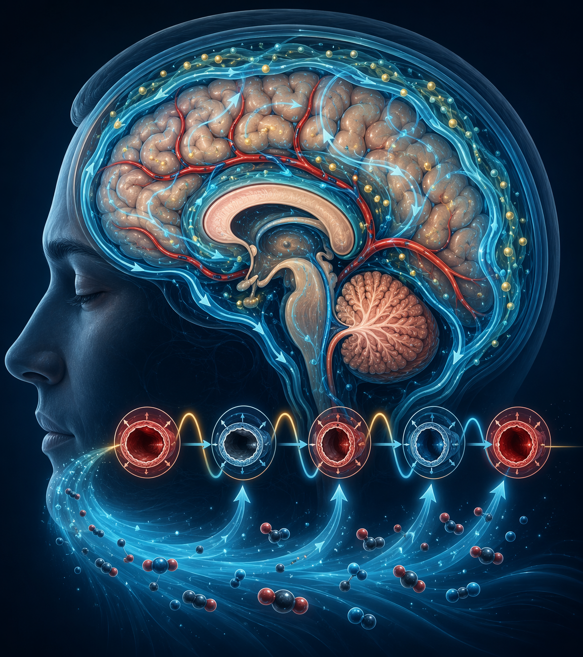
After the session, blood measurements showed increased beta-amyloid in all 12 participants. That is the finding behind the headline: more amyloid in blood after the intervention is interpreted as a possible sign that amyloid moved from the brain into circulation.[1][2]
The tau result was narrower. Tau clearance was observed in the four participants who either had elevated baseline tau or mild cognitive impairment. It was not reported as a uniform effect across all 12 people.[1] That distinction is easy to lose in a headline, but it changes the claim. The study does not show that one CO₂ session broadly clears tau in everyone. It suggests that when tau is already elevated or cognition is already impaired, the same vascular-fluid mechanism may be able to move measurable tau as well.
Researchers also reported no side effects in this small group.[1] That is reassuring for a supervised study visit, not a safety certificate for unsupervised use. Twelve people is not enough to detect uncommon risks, and a conference presentation is not the same as a peer-reviewed clinical trial.
The most sobering detail is the time course. The biomarker changes returned to baseline within about an hour after the session.[1] Whatever happened was temporary. That does not make the result meaningless; temporary biological effects can be scientifically revealing. But it does mean the evidence does not support claims of durable disease modification, prevention, or treatment.
Why CO₂ can imitate part of deep sleep
Carbon dioxide is a powerful signal to blood vessels. When CO₂ rises in the body, cerebral blood vessels can dilate. When it falls back, they constrict. If that rise and fall is repeated in a controlled cycle, the vessels do not just change diameter once; they pulse.
During deep, slow-wave sleep, the brain shows slow vascular and fluid rhythms that are thought to help move cerebrospinal fluid through brain tissue. This waste-clearance pathway is commonly called the glymphatic system. Foundational animal research helped establish that this system is much more active during sleep than wakefulness, and later work has connected slow-wave sleep, vasomotion, and clearance of molecules from the brain.[1][2]
The CO₂ intervention is compelling because it reaches for the same physical lever. It does not ask neurons to “sleep.” It asks blood vessels to reproduce a sleep-like rhythm strongly enough to push fluid movement. If proteins then rise in the blood afterward, the simplest mechanistic interpretation is that the intervention has increased movement from brain tissue toward circulation.
That interpretation still has limits. Blood biomarkers are indirect. They can suggest clearance, but they do not show every step of the journey from brain tissue to cerebrospinal fluid to blood. Human glymphatic biology is also still being characterized; much of the strongest mechanistic foundation began in rodent models. The bridge to humans is plausible, not complete.
Amyloid is not the whole story
The Alzheimer’s-associated proteins are what made the July 2026 study newsworthy, but they may not be the only useful readout of this mechanism. A 2025 study published in npj Parkinson’s Disease reported that a related CO₂-driven approach enhanced clearance of alpha-synuclein, the protein closely associated with Parkinson’s disease.[3]
That does not mean CO₂ is a treatment for Parkinson’s disease either. It means the mechanism may be broader than one protein. If rhythmic vessel motion increases fluid movement through brain tissue, it would make sense that more than one waste product could be affected. The value of the Parkinson’s work is that it supports the clearance mechanism without turning the Alzheimer’s result into a miracle story.
Why this is not the same as a stuffy room or sleep apnea
The paradox is real enough to be confusing: CO₂ is often discussed as a problem in sleep, yet here it is being used to imitate a helpful sleep-linked rhythm. The difference is exposure pattern and control.
Poor ventilation, sleep-disordered breathing, and chronically abnormal blood gases are not the same as alternating room-air-level CO₂ with 5% CO₂ every 35 seconds through medical equipment for 30 minutes. One is uncontrolled physiology during sleep or in an indoor environment. The other is a deliberately timed stimulus designed to produce vascular oscillation under supervision.[1][2]
This is exactly where the home-experiment temptation becomes dangerous. A bag, a mask, a canister, or an improvised breathing setup cannot reproduce a supervised protocol with monitored gas concentrations. High-dose CO₂ can be risky. The fact that 5% CO₂ was used in a research setting does not make 5% CO₂ a wellness tool.
The better way to read the study is not “CO₂ is good.” It is “timed vascular motion may matter.” That is a narrower claim, and it is the one the evidence can actually carry.
What, if anything, is actionable now?
There is no consumer version of this procedure to recommend. The Alzheimer’s-specific data involve 12 participants, a short monitored exposure, post-session biomarker changes, and results that returned to baseline within about an hour.[1][2] That is the territory of hypothesis testing, not self-care.
Researchers are also interested in whether slower abdominal breathing practices, such as those used in yoga or meditation, might mildly raise CO₂ enough to influence glymphatic activity.[1] That is a much lower-risk area of investigation, but it should not be oversold either. A calming breathing practice is not equivalent to breathing 5% CO₂ through a mask in a laboratory.
The practical takeaway narrows back to sleep itself. The experiment is exciting because it shows how physically active deep sleep may be: blood vessels pulsing, fluid shifting, waste products moving. Sleep is not simply a pause in brain activity. It is a different operating mode, and one of its jobs may be maintenance that waking life cannot easily replace. The study gives researchers a possible way to probe brain clearance on demand. For everyone else, it is a reminder that deep sleep is not decorative. It is part of the machinery.
References
- Inhaling high-dose CO2 clears Alzheimer’s proteins from the brain, New Scientist.
- Researchers Study Whether Intentionally Manipulating Blood Carbon Dioxide Levels Might Enhance Brain Health, UNM HSC Newsroom.
- Breathing CO2 May Help the Brain Clear Toxic Proteins in Parkinson's Disease, Touro University.
Comments
Join the discussion with an anonymous comment.