
Carbon dioxide is not an obvious candidate for a brain-health headline. Most people who follow sleep research have learned to worry about it: stuffy bedrooms, untreated sleep apnea, impaired breathing, poor sleep architecture. So the recent claim that inhaling CO2 might help move Alzheimer’s-related proteins out of the brain deserves a careful pause, not a leap.
The key word is not CO2. It is rhythm.
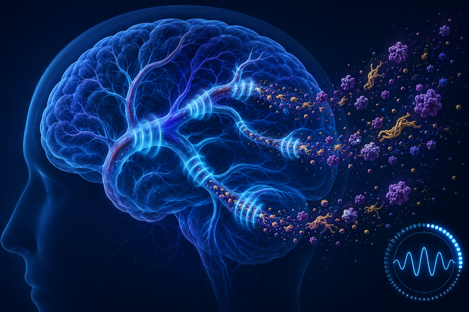
At the Alzheimer’s Association International Conference in London in July 2026, Sephira Ryman presented early human data in which 12 participants inhaled 5% CO2 in 35-second ON/OFF cycles for 30 minutes. The group included eight healthy older adults and four people with elevated tau and mild cognitive impairment. Blood amyloid-beta rose within 15 minutes after the session, tau rose in the MCI subgroup, no adverse events were reported, and the measured effects returned to baseline about an hour after the intervention.[1]
That is interesting. It is also very small, very early, and very temporary. The study does not show that CO2 therapy prevents Alzheimer’s disease, slows cognitive decline, improves memory, or safely belongs in anyone’s home. What it suggests is narrower and still important: brief, controlled CO2 oscillation may be able to trigger a vascular pumping pattern that resembles one part of deep sleep’s normal cleaning physiology.
What Actually Happened in the CO2 Session
The intervention was not ordinary breathing into a bag. It was not sitting in a high-CO2 room. It was not a wellness “detox.” Participants received a controlled gas mixture: 5% CO2, delivered intermittently, with 35 seconds on and 35 seconds off, for a total session of 30 minutes.[1]
| Feature | What was reported |
|---|---|
| Participants | 12 total: 8 healthy older adults and 4 with elevated tau/MCI |
| CO2 dose | 5% CO2 |
| Pattern | 35-second ON/OFF cycles |
| Session length | 30 minutes |
| Observed blood markers | Amyloid-beta rose after the session; tau rose in the MCI subgroup |
| Safety report | No adverse events in this small supervised group |
| Duration of effect | Returned to baseline about 1 hour after intervention |
Those details matter because dose and timing are the experiment. If the claim is reduced to “CO2 clears Alzheimer’s proteins,” the most important part disappears. The proposed effect depends on repeatedly raising and lowering CO2, not simply increasing CO2 exposure.
The blood-marker change also needs careful interpretation. A rise in amyloid-beta or tau in blood after a session may mean more of those proteins moved from brain-related compartments into circulation. It does not by itself prove that the brain has been protected, that harmful deposits have been reduced, or that symptoms will change. A biomarker can be a signal of movement without being proof of clinical benefit.
The Sleep Connection: A Pump, Not a Metaphor
The reason this CO2 finding belongs in sleep science is the glymphatic system: the brain’s fluid-clearance pathway. During non-REM slow-wave sleep, rhythmic changes in blood-vessel diameter help drive cerebrospinal fluid, or CSF, into brain tissue. That incoming fluid can help carry away waste proteins, including amyloid-beta and tau.
The useful image is not a sponge being squeezed once. It is closer to a pulse-driven exchange. Blood vessels widen and narrow. CSF shifts through spaces around those vessels and through brain tissue. Waste-related proteins can be carried away from the compartments where they accumulate and eventually appear downstream in blood.
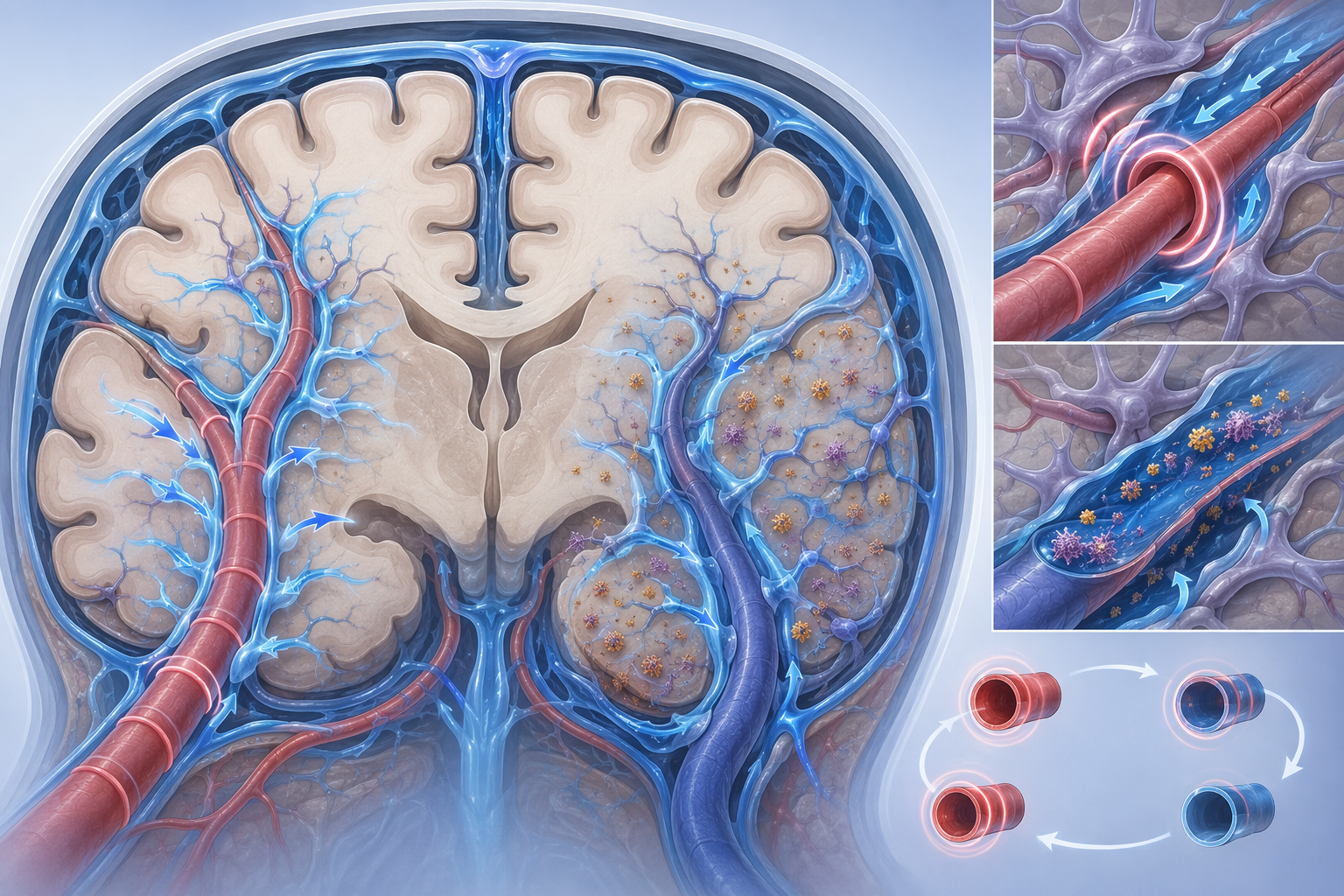
CO2 has a strong effect on cerebral blood vessels. When CO2 rises, those vessels tend to dilate. When CO2 falls back, they narrow toward baseline. In an intermittent protocol, that repeated change can create vasomotion: a widening-and-narrowing rhythm that may push CSF in a way that resembles part of what happens naturally during deep sleep.
That is the mechanically elegant part of the study. It does not require pretending that CO2 is broadly “good” for the brain. It proposes that a controlled oscillation in CO2 can act as a temporary vascular metronome.
The Alzheimer’s presentation was not the first time this research direction appeared. A November 2025 study in npj Parkinson’s Disease reported that intermittent hypercapnia cleared alpha-synuclein in people with Parkinson’s disease and in healthy older adults.[2] Alpha-synuclein is a different protein and Parkinson’s is a different disease, so the result should not be imported wholesale into Alzheimer’s. But it supports the broader idea that CO2-driven vascular rhythms may be able to move neurodegeneration-related proteins, at least transiently, under controlled conditions.
Why “Intermittent” Changes the Meaning of CO2
A 35-second cycle is not a decorative detail. It separates the research question from a dangerous home experiment.
Intermittent hypercapnia means CO2 rises and falls in a controlled pattern while people are monitored. Sustained CO2 exposure means the gas remains elevated. Those are biologically different situations. One is being studied as a way to generate vascular motion. The other can signal inadequate ventilation, disordered breathing, or an unsafe environment.
This distinction is especially important for sleep. If CO2 simply improved brain clearance because “more is better,” then high-CO2 sleep environments would look helpful. They do not. In a 2021 Indoor Air study, sleep quality declined linearly as indoor CO2 rose from 800 to 3,000 ppm, and slow-wave sleep decreased proportionally.[3]
That finding should make the safety boundary easier to understand. Slow-wave sleep is the stage most relevant to glymphatic clearance. Sustained indoor CO2 increases were associated with less of it, not more.[3] So the sleep lesson is not to tolerate stale air or seek out CO2 exposure. It is almost the opposite: protect deep, unobstructed sleep, and do not confuse a laboratory CO2 rhythm with chronically poor ventilation.
The Safety Boundary Is Part of the Science
High CO2 is not a supplement. It changes breathing drive, blood chemistry, vascular tone, and oxygen-CO2 balance. In the New Scientist report, Ryman and independent experts Nick Fox of University College London and Jeffrey Iliff of the University of Washington warned that high CO2 concentrations can be dangerous and life-threatening outside controlled clinical settings.[1]
That warning is not legal padding. It is central to interpreting the result. The same physiological lever that makes CO2 interesting—its ability to alter cerebral blood vessels and breathing chemistry—is what makes unsupervised exposure risky.
A supervised research setup can control the gas mixture, timing, delivery system, participant selection, monitoring, stopping rules, and emergency response. A home setup cannot reproduce those safeguards by improvisation. Even if someone could copy the headline number, the missing clinical context would still be the hazard.
- The reported Alzheimer’s session used a specific 5% CO2 mixture, not an unknown concentration.
- The exposure alternated on and off every 35 seconds, rather than remaining continuously elevated.
- The session lasted 30 minutes, with post-session measurement.
- The participants were observed in a research context, not self-experimenting.
- The absence of adverse events came from only 12 people, which cannot establish broad safety.
The small sample matters here. “No adverse events” in 12 supervised participants is reassuring enough to justify further research. It is not large enough to reassure the general public, older adults with cardiovascular or pulmonary disease, people with sleep apnea, or anyone tempted to manipulate breathing gases on their own.
What the Biomarker Rise Can and Cannot Mean
For families worried about Alzheimer’s, the emotional pull is obvious. Amyloid-beta and tau are not abstract names once someone in the family has mild cognitive impairment or a diagnosis. A visible shift in those markers feels like action.
But the result reported so far is movement, not outcome. Amyloid-beta rose in blood after the session, and tau rose in the MCI subgroup.[1] The simplest interpretation is that the intervention may have increased transfer of these proteins out of brain-related compartments into circulation. The study did not show that the total brain burden of pathology meaningfully fell. It did not show that repeated sessions are beneficial. It did not show that cognition changes.
The return to baseline after about an hour is one of the most important facts in the whole story.[1] A short-lived biomarker change can still be scientifically valuable; it may tell researchers that the pump can be engaged. But it is a long distance from a one-hour physiological signal to a disease-modifying treatment.
Future studies would need to answer harder questions: whether repeated supervised sessions are safe, whether protein movement changes brain measures over time, whether MCI patients differ from healthy older adults, whether there are cardiovascular or respiratory exclusions, and whether any cognitive or functional outcome improves. Without those answers, the honest category is experimental mechanism, not therapy.
Why Researchers Are Pursuing This Anyway
The University of New Mexico Health Sciences program has described research into whether intentionally manipulating blood CO2 levels might enhance brain health.[4] That broader context matters because the Alzheimer’s presentation is not merely a one-off curiosity. It sits inside an active attempt to understand whether vascular and respiratory physiology can be used to influence brain-fluid movement.
That research direction is plausible because the brain’s clearance system is already tied to sleep, vascular pulsation, breathing, and CSF flow. It is also difficult because each of those systems can become unsafe when pushed too hard. A mechanism can be real and still not be ready for use.
The Parkinson’s alpha-synuclein finding strengthens the mechanistic case without settling the clinical one.[2] It suggests that intermittent hypercapnia may be acting on a general clearance pathway rather than a single Alzheimer’s marker. Still, protein movement across conditions is not the same as treating either condition. The burden of proof becomes higher, not lower, when a mechanism starts to look broadly powerful.
What About Breathing Practices?
There is a safer, much milder research-adjacent question: can slow breathing practices influence CO2 enough to affect glymphatic function? A 2026 Journal of Cerebral Blood Flow & Metabolism review discussed how yoga or meditative breathing may modestly modulate CO2 and potentially stimulate glymphatic activity.[5]
That is a perspective-level idea, not proof that breathing exercises clear Alzheimer’s proteins. No material here supports the claim that yoga lowers amyloid, removes tau, or substitutes for clinical treatment. Slow abdominal breathing may be reasonable for relaxation or sleep preparation for many people, but it should not be marketed as a home version of intermittent 5% CO2 therapy.
The difference is scale and control. Gentle breathing practices work within ordinary physiology. The CO2 protocol deliberately alters inhaled gas concentration under supervision. Treating them as interchangeable would erase the very safety distinction that makes the experimental protocol interpretable.
The Practical Answer for Now
If the question is whether inhaling CO2 can clear Alzheimer’s-related proteins from the brain, the most accurate answer is: in a tiny supervised study, intermittent 5% CO2 in 35-second ON/OFF cycles temporarily increased blood markers consistent with movement of amyloid-beta, and tau in the MCI subgroup.[1] That is a real signal worth studying.
If the question is whether CO2 therapy is safe for Alzheimer’s prevention, the answer is no. Not outside a clinical research setting. The human evidence is preliminary, the measured effect was transient, and experts have warned that high CO2 can be dangerous and life-threatening when not carefully controlled.[1]
The sleep lesson is more durable than the headline. Deep, healthy slow-wave sleep already performs part of this clearance work through rhythmic vascular and CSF movement. CO2 therapy is interesting because it may imitate one piece of that physiology for a short time. It is not a reason to experiment with gas exposure, tolerate poor air, or treat “brain detox” as a household project.
References
- Inhaling high-dose CO2 clears Alzheimer's proteins from the brain, New Scientist.
- Intermittent hypercapnia clears alpha-synuclein in Parkinson's and healthy older adults, npj Parkinson's Disease, November 2025.
- Sleep quality and sleep structure in relation to indoor carbon dioxide concentration, Indoor Air, 2021.
- Researchers study whether intentionally manipulating blood carbon dioxide levels might enhance brain health, UNM Health Sciences.
- Yoga and meditative breathing as modulators of glymphatic function, Journal of Cerebral Blood Flow & Metabolism, 2026.
Comments
Join the discussion with an anonymous comment.