
The clean version of the story is easy to remember: during sleep, the brain washes away amyloid beta and tau, two proteins linked to Alzheimer’s disease. The harder version is the one worth keeping. Sleep is strongly tied to lower brain concentrations of these proteins, but scientists are still arguing over the route by which that happens. Does sleep actively clear amyloid beta and tau from the brain, or does wakefulness simply generate more of them, making sleep look like a cleanup shift by comparison?
That distinction may sound like a technical quarrel until it reaches the clinic. If clearance is the dominant mechanism, then therapies that improve fluid movement through brain tissue become more attractive. If production is the dominant mechanism, then the better target may be the neuronal activity of wakefulness that drives protein release. For an individual trying to protect brain health, though, the immediate advice does not swing wildly with the answer. Both sides of the dispute agree that sleep meaningfully affects Alzheimer’s-linked proteins and that poor sleep is associated with higher dementia risk over time.
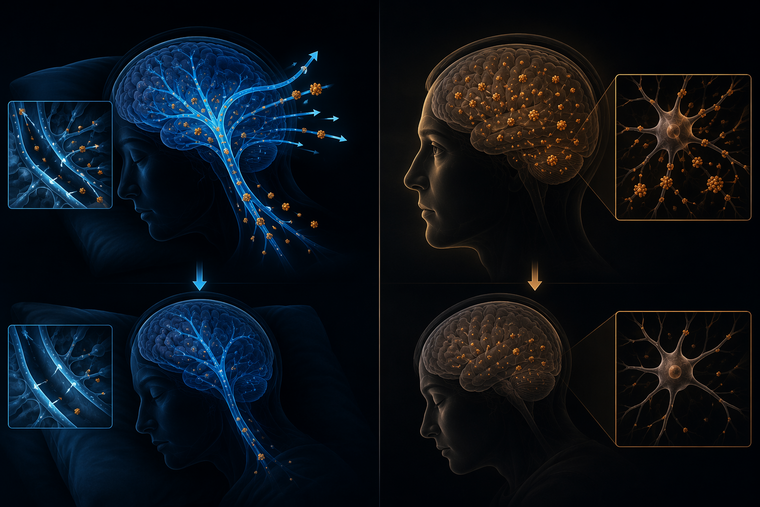
Why the “brain washes itself at night” idea became so persuasive
The popular clearance story traces largely to a 2013 mouse study from Maiken Nedergaard’s group. The experiment used a tracer injected into the cisterna magna, a cerebrospinal-fluid-filled space at the base of the brain, to watch how fluid moved through the brain during wakefulness, sleep, and anesthesia. In that work, clearance from the brain was reported to be about 60% higher during sleep and anesthesia than during wakefulness.[1]
The study was vivid for a reason. It did not merely report a correlation between bad sleep and later disease. It offered a physical model: cerebrospinal fluid enters along spaces around blood vessels, exchanges with interstitial fluid inside brain tissue, and helps carry away soluble waste. The group called this system “glymphatic,” a name meant to evoke lymphatic drainage while acknowledging the role of glial cells in the brain.[1]
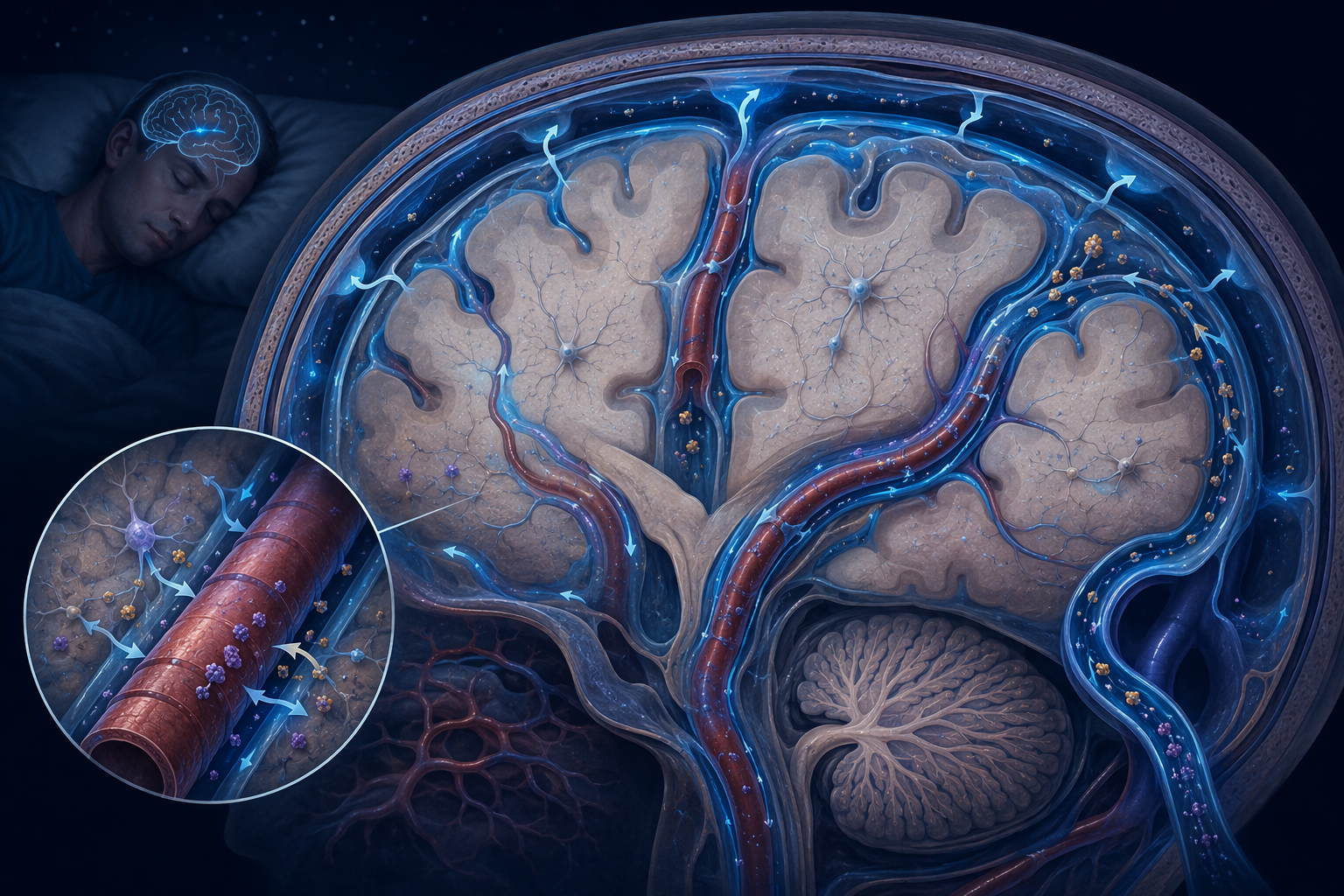
It is not hard to see why the image traveled. Sleep, a state that can feel passive from the outside, suddenly had visible work to do. The brain was not idling; it was maintaining itself. In a field where Alzheimer’s disease is often described through slow accumulation and delayed symptoms, the idea that nightly sleep might alter the handling of amyloid beta gave the public a mechanism it could picture.
But the study did not prove every version of the slogan later attached to it. It was a mouse experiment. It used a particular route of tracer delivery. It measured movement of tracer and clearance under defined experimental conditions. It supported the glymphatic model; it did not, by itself, establish that normal human sleep reliably rinses Alzheimer’s pathology out of the brain in the way a household drain clears a sink.
Clearance and production can look similar from a distance
A lower concentration of amyloid beta or tau after sleep can be explained in more than one way. One explanation is clearance: the protein is made, then removed more efficiently during sleep. Another is production: the waking brain releases or generates more of the protein, and sleep reduces that production. The measured level drops either way, but the biology is not the same.
| Observation | Clearance explanation | Production explanation |
|---|---|---|
| Alzheimer’s-linked proteins are lower after sleep | Sleep increases removal from brain tissue or fluid compartments | Wakefulness increases generation or release, while sleep slows it |
| Sleep deprivation raises measured protein levels | Less sleep means less removal time or less efficient transport | More wakefulness means more neuronal activity and more protein production |
| A therapy target is chosen | Improve glymphatic or fluid-transport function | Reduce wake-related synaptic drivers of protein release |
This is where many public explanations lose their footing. They move from “sleep changes levels of Alzheimer’s-linked proteins” to “sleep clears those proteins” without pausing over the measurement. A concentration is an outcome of inputs and outputs. To know whether clearance increased, the method has to capture removal, not merely the final amount.
That does not make the clearance idea flimsy. It makes it specific. The glymphatic model is a claim about fluid movement, exchange, and export. It deserves to be tested with methods that can distinguish movement into brain tissue, movement out of brain tissue, and changes in protein production during wake and sleep.
The 2024 challenge did not simply “debunk” the mouse work
In 2024, a group led by Nicholas Franks challenged the prevailing interpretation with another mouse experiment. Instead of injecting tracers into the cisterna magna, the researchers injected tracers directly into the brain parenchyma and measured how they cleared from that local site. They reported that clearance was reduced during sleep and anesthesia, the opposite direction from the 2013 glymphatic result.[2]
That finding forced a useful slowdown. The two experiments were not interchangeable versions of the same measurement. Nedergaard’s side argued that the Franks method measures efflux from a localized injection point rather than network-level glymphatic influx from cerebrospinal fluid into the brain. Franks argued that his approach is a more direct test of whether material leaves brain tissue. Both sides acknowledged that the other technique measures something real; the disagreement is over which measurement answers the clearance question readers think they are asking.[2]
That is a less satisfying headline than “sleep cleans the brain” or “sleep cleaning theory collapses,” but it is a better description of the dispute. A tracer introduced at the base of the brain and a tracer placed directly into tissue may travel through different compartments, disturb tissue differently, and emphasize different parts of fluid handling. One can be informative about large-scale influx while the other is informative about local efflux. Neither automatically cancels the other.
The methodological fight also protects against a common mistake in translating animal studies. Mice allow invasive measurements that are impossible or unethical in healthy humans. That is why they matter. It is also why they can be overextended. The more precise conclusion is that sleep-state changes in fluid and solute movement have been observed in mouse experiments, but the direction and meaning of “clearance” depend heavily on how the experiment defines and measures it.
The 2026 human trial adds weight, not a final verdict
The strongest new reason to take the clearance model seriously in humans is a 2026 randomized crossover trial by Dagum and colleagues in Nature Communications. The study included 39 participants and compared normal sleep with sleep deprivation. After normal sleep, morning plasma levels of Aβ40, Aβ42, np-tau181, np-tau217, and p-tau181 were higher than after sleep deprivation.[3]
At first glance, higher blood levels can sound backwards. If these are Alzheimer’s-linked proteins, why would an increase after sleep be encouraging? The logic is compartmental. Plasma is not brain tissue. A rise in blood after sleep can be interpreted as evidence that proteins moved out of the brain and into peripheral circulation. That makes the study more directly relevant to clearance than a simple brain-level measurement would be.
The trial also used a neuro-glymphatic model that explained 50% to more than 90% of overnight variance in the measured biomarkers, depending on the marker and model specification.[3] That is the kind of result that deserves attention: randomized crossover human evidence, sleep manipulated rather than merely observed, and multiple Alzheimer’s-related biomarkers moving in a direction consistent with sleep-driven export.
It also deserves the ordinary cautions that often disappear when a result is appealing. The sample was small. The finding has not yet been independently replicated at scale. The study was funded by Applied Cognition, a company developing glymphatic-targeting therapeutics.[3] Commercial funding does not invalidate a result, but it does raise the value of independent confirmation, especially when the result supports a therapeutic direction in which the funder has an interest.
So the 2026 trial changes the posture of the debate without ending it. Before this kind of human evidence, the clearance story leaned heavily on animal mechanisms and indirect human inference. Now there is causal human evidence consistent with sleep-driven glymphatic clearance. That is a meaningful shift. It is not the same as knowing the size of the effect in older adults, in people with sleep apnea, in people with established Alzheimer’s pathology, or across years of repeated sleep disruption.
What can be safely believed now
The safest belief is narrower than the most popular story and stronger than the most dismissive one. Sleep affects amyloid beta and tau biology. Sleep deprivation and poor sleep are not neutral states for the brain. The mechanism probably includes both reduced production during sleep and some form of clearance, with the balance varying by brain region, protein type, disease stage, and timescale.
It is also safe to say that “clearance” is not one measurement. Clearing a tracer from a local injection site, moving cerebrospinal fluid into perivascular spaces, exporting proteins into blood, and lowering tissue concentration are related questions, not identical ones. When two studies appear to disagree, the first question should be what each one actually measured.
What is not safe is turning the glymphatic hypothesis into a finished public-health certainty. The human evidence is promising but early. The rodent evidence remains important but method-dependent. The 2024 challenge is serious but not a demolition. Anyone claiming a clean winner is smoothing over the part of the science that still needs work.
What to do while the mechanism is still being worked out
The practical conclusion is less conflicted than the mechanism. Protecting sleep, especially consolidated, high-quality sleep, remains a sensible dementia-risk strategy because both interpretations point in the same direction. If sleep clears proteins, then deep sleep may support removal. If wakefulness drives production, then chronic sleep loss extends the period in which those proteins are produced or released. Either way, treating sleep as optional is hard to defend.
- Prioritize regular, sufficient sleep rather than treating “detox sleep” as an occasional recovery ritual.
- Take symptoms of sleep apnea seriously, especially loud snoring, witnessed breathing pauses, morning headaches, or excessive daytime sleepiness.
- Avoid alcohol before bed when possible, because it can degrade sleep quality even when it makes falling asleep feel easier.
- Be cautious with products that claim to boost glymphatic flow before the human evidence has been independently replicated.
Those recommendations do not require pretending that every protein molecule is being washed away on a nightly schedule. They rest on the broader, better-supported point: sleep is one of the modifiable behaviors most consistently tied to brain protein regulation and dementia risk. The machinery is still being mapped. The need to protect it is not waiting for the map to be perfect.
References
- The Brain’s Night Shift, University of Rochester Medical Center, The Brain's Night Shift
- The great brain clearance and dementia debate, Nature, The great brain clearance and dementia debate
- Dagum et al., Nature Communications, s41467-026-68374-8
Comments
Join the discussion with an anonymous comment.