
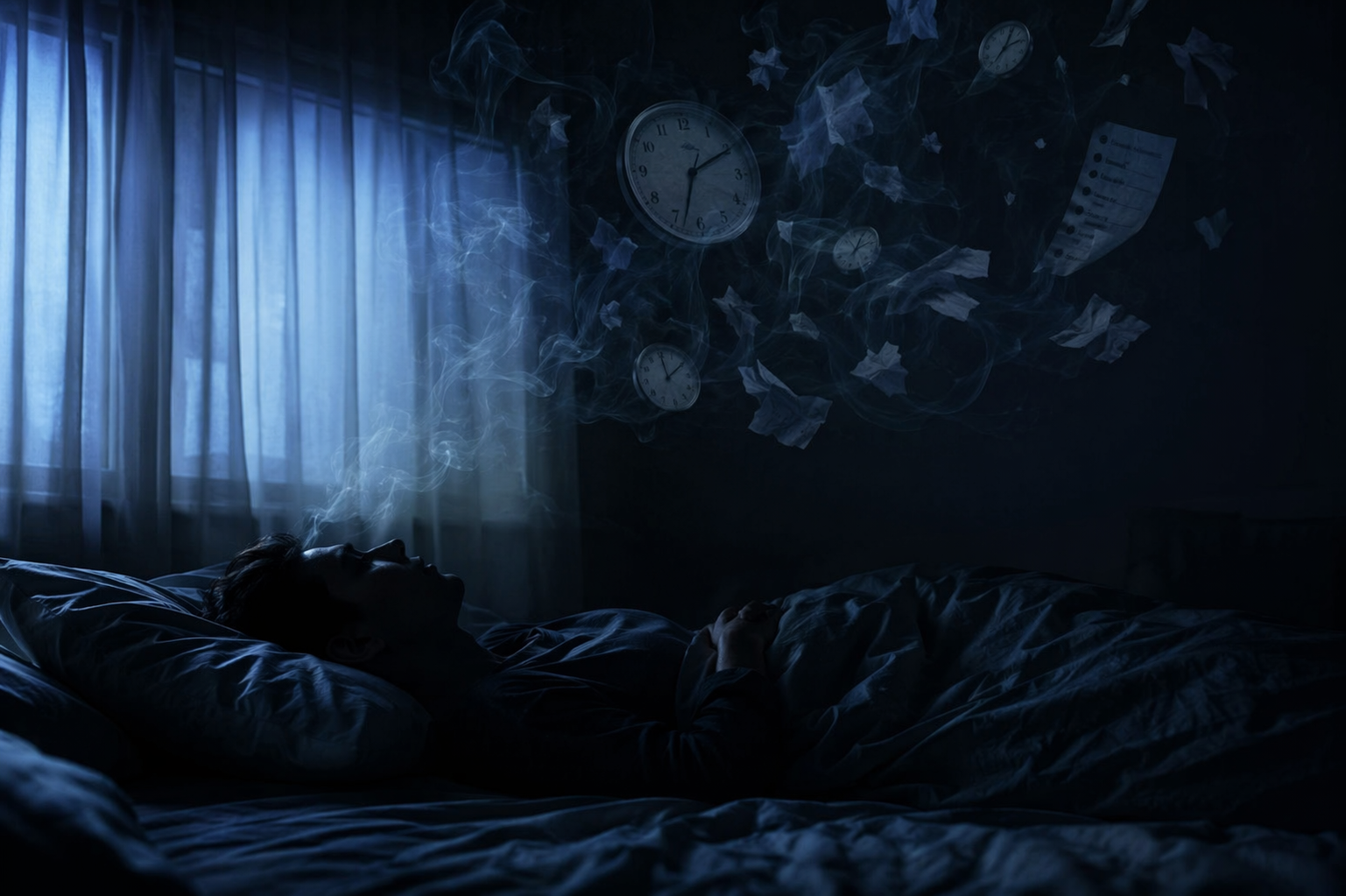
After a concussion or traumatic brain injury, “sleep better” can sound like sensible advice and still miss the problem. Many people are not simply staying up too late, scrolling too long, or forgetting the rules of a calm bedtime. Their sleep may have become unstable, mistimed, unusually heavy, strangely light, or disconnected from the tiredness they feel during the day.
That mismatch is common enough to deserve more precision. A meta-analysis by Mathias and Alvaro found that sleep disturbances, disorders, and sleep-related problems affect an estimated 30% to 70% of people after traumatic brain injury, roughly three times the rate seen in the general population.[1] But prevalence is only the doorway. The more useful question is what kind of sleep-control system was disrupted.
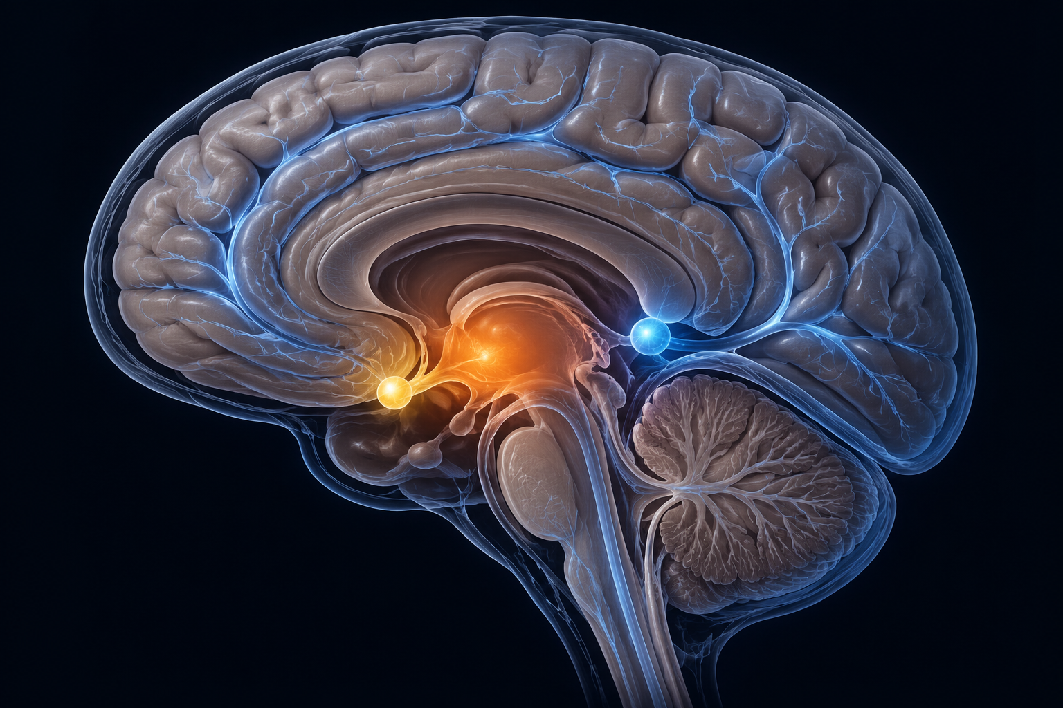
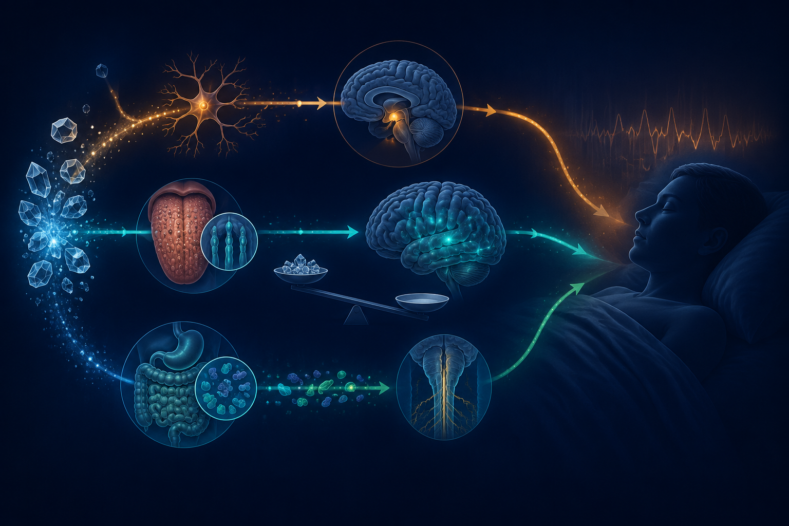
Sleep patterns during brain injury recovery can change through more than one biological route. The strongest evidence points to at least four systems: the orexin/hypocretin arousal network, the suprachiasmatic circadian clock, pineal melatonin signaling, and sleep-linked glymphatic waste clearance. These systems do not produce one neat disorder. They produce overlapping complaints that can look like insomnia, hypersomnia, delayed sleep phase, fragmented sleep, or persistent daytime sleepiness.
The Arousal System Can Be Injured, Not Just Overstimulated
Orexin, also called hypocretin, is one of the brain’s wake-stabilizing signals. It is produced by neurons in the lateral hypothalamus and helps keep the sleep-wake switch from drifting at the wrong time. When this system is healthy, it supports sustained wakefulness during the day and more coherent boundaries between being awake and asleep.
This is where post-TBI sleep stops fitting ordinary insomnia language. In a 2007 study discussed in a later synthesis by Sandsmark, Elliott, and Lim, Baumann and colleagues measured cerebrospinal fluid orexin in 44 acute moderate-to-severe TBI patients and found low levels in 95% of them. The same body of work also includes autopsy evidence showing loss of orexin-producing neurons in the lateral hypothalamus after TBI.[2]
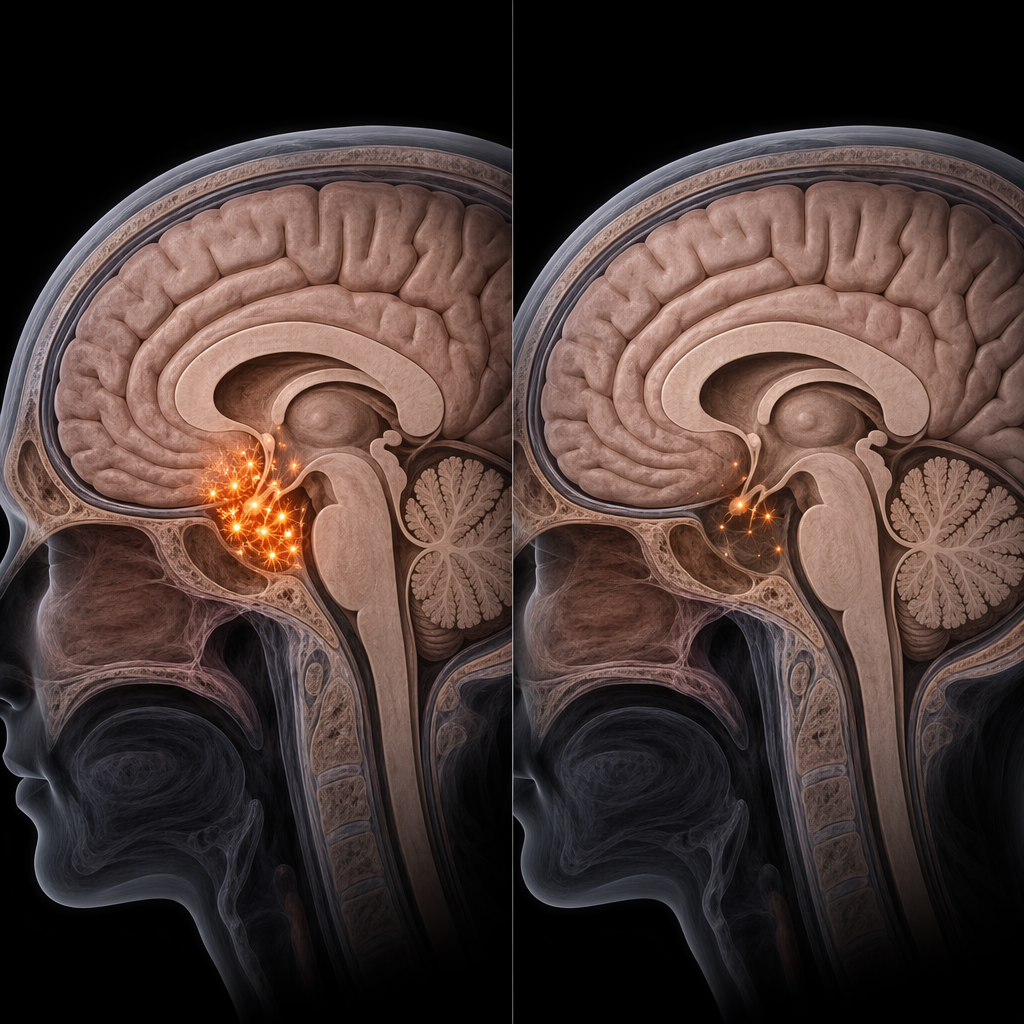
That finding does not mean every person with a mild concussion has orexin deficiency. The 95% figure came from a single study of acute moderate-to-severe TBI patients, not a large, repeated sample across all injury severities.[2] It also does not turn every post-TBI sleep complaint into narcolepsy. What it does show, very plainly, is that traumatic injury can affect the machinery that stabilizes wakefulness itself.
The symptom pattern that follows may not be “I cannot fall asleep because I am anxious.” It may be “I sleep for hours and still feel drugged,” “I cannot stay alert reliably,” or “my sleep and wakefulness no longer hold their shape.” A person with that pattern can follow every item on a sleep hygiene fundamentals checklist and still be dealing with a damaged arousal pathway.
This is why the first clinical distinction matters: is the dominant complaint trouble initiating sleep, repeated awakenings, excessive daytime sleepiness, long sleep time, poor timing, or unrefreshing sleep? A generic insomnia label collapses those differences. Orexin biology makes the collapse especially costly, because an arousal-system problem asks a different question than bedtime discipline does.
The Brain Clock May Shift the Night Later
The circadian system is the brain’s timing architecture. Its central coordinator, the suprachiasmatic nucleus, uses light information and clock-gene rhythms to help schedule alertness, body temperature, hormone release, and sleep pressure across the 24-hour day. A person can be exhausted and still not be biologically ready for sleep if that timing system is delayed.
For readers who want the broader biology first, the basic machinery is covered in circadian rhythm mechanisms. After TBI, the concern is that this machinery may not simply be ignored by poor habits; it may be dysregulated by injury.
Kuo and colleagues reported dysregulation of circadian clock genes, including Per2, Cry1, and Bmal1, in peripheral blood leukocytes after TBI. The same 2024 work reported disrupted brain temperature rhythms in 40.7% of moderate-to-severe patients, a signal that the timing system’s normal daily pattern can be disturbed after injury.[3]
That matters because circadian disruption often masquerades as bad sleep behavior. If someone’s internal night has moved later, a 10:00 p.m. bedtime may be a demand to sleep at the wrong biological time. The person lies there awake, then sleeps better closer to morning, then looks unmotivated or noncompliant when they cannot wake easily for work, school, therapy, or family care.
Ayalon and colleagues documented circadian rhythm sleep disorders after mild traumatic brain injury, especially delayed sleep phase, and noted that these cases may be misdiagnosed as insomnia.[4] That is a small but clinically important distinction. Insomnia treatment often tries to reduce conditioned arousal around bed. Delayed sleep phase asks whether the sleep window itself has moved.
This is the post-concussion patient who is told to “go to bed earlier” and then fails the assignment night after night. The failure may not be a character trait. It may be a timing problem. The practical next question is not whether they understand bedtime; it is whether their sleep onset, wake time, light exposure, body temperature rhythm, and daytime alertness all point to a shifted clock.
A more precise label changes the pathway. Someone whose pattern resembles delayed sleep phase syndrome may need carefully timed morning light, evening light reduction, schedule anchoring, and sometimes clinician-guided chronobiotic strategies. The logic is closer to circadian treatment, including approaches used in conditions such as shift work sleep disorder, than to a generic reminder to relax before bed.
Melatonin Findings Point in the Same Direction, With Caution
Melatonin is not a sedative switch. It is a darkness signal, produced through the pineal pathway, that helps communicate biological night to the rest of the body. In ordinary insomnia, melatonin supplements are often oversold because many people’s main problem is not a missing melatonin signal. After TBI, the picture may be different for some patients, but the evidence still needs careful handling.
Grima and colleagues studied 23 chronic TBI patients and found that they produced 42% less salivary melatonin than matched controls, along with a delay in melatonin onset.[5] The number is striking, but the sample is small. It supports the possibility that melatonin signaling may be reduced or mistimed after TBI; it does not prove that every post-TBI sleep problem is a melatonin deficiency.
The more useful takeaway is about fit. If a person’s sleep pattern suggests circadian delay, then melatonin timing may become relevant in a way that differs from routine insomnia self-treatment. Timing, dose, and clinical context matter. A late, high-dose supplement can create a different set of problems than a carefully timed circadian cue.
Readers comparing this with general supplement advice should separate two questions: when melatonin actually works for insomnia, and whether a brain injury has shifted or weakened the body’s night signal. Those are related questions, not the same question.
Sleep May Also Affect Brain Cleanup, But the Evidence Is Not a Repair Button
The glymphatic system is often described as the brain’s waste-clearance pathway. It is most active during sleep and is thought to help move fluid through perivascular spaces, clearing metabolic byproducts from brain tissue. After TBI, this system is interesting because recovery is not only about getting unconscious for enough hours. Sleep quality may influence the internal environment in which the injured brain is trying to stabilize.
An OHSU MRI study reported that poor sleep after traumatic brain injury was linked with enlarged perivascular spaces and more post-concussive symptoms, a pattern the investigators interpreted as suggesting a role for impaired glymphatic clearance during sleep.[6] The careful word is linked. This kind of finding does not prove that poor sleep directly causes enlarged perivascular spaces, or that improving sleep alone reverses the imaging pattern.
Still, the association helps explain why sleep after TBI can feel consequential in a way ordinary tiredness does not. Fragmented, mistimed, or shallow sleep may not give the brain the same recovery conditions as consolidated sleep. The science is not ready to turn glymphatic clearance into a simple consumer promise, but it is strong enough to make dismissive advice feel inadequate.
Why a Sleep Hygiene Sheet Often Feels Like the Wrong Tool
Sleep hygiene is not useless. A stable wake time, morning light, lower evening light, regular meals, reduced alcohol, and a bedroom that is cool and quiet can support recovery. These steps reduce avoidable friction around sleep, and they may be especially helpful when the nervous system is already strained.
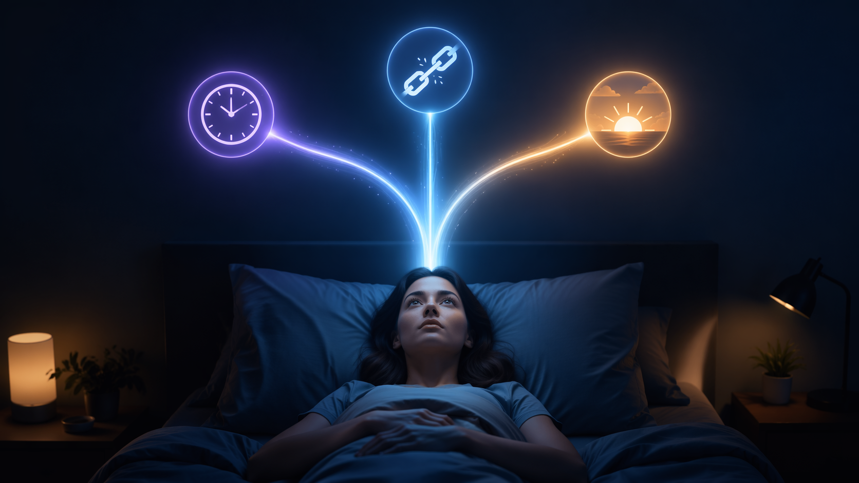
The problem is sequencing. If the first and only response to post-TBI sleep disruption is “avoid caffeine and screens,” the care plan may skip the diagnostic work. The same complaint — “I can’t sleep” — can represent at least four different situations:
- Arousal instability: the person cannot maintain normal wakefulness or feels profoundly sleepy despite long sleep.
- Circadian delay: the person can sleep, but only at a later biological time than their schedule allows.
- Melatonin mistiming or reduction: the darkness signal may be delayed, blunted, or poorly aligned with the desired sleep window.
- Fragmented recovery sleep: sleep may be disturbed in a way that correlates with symptom burden and possibly impaired waste-clearance physiology.
This is also where evidence-based insomnia care needs a fair distinction. CBT-I is a real treatment for chronic insomnia, not a wellness slogan. But even CBT-I works best when the clinical target is correct. A person with delayed sleep phase after mild TBI may need circadian evaluation before sleep restriction is intensified. A person with disabling daytime sleepiness after moderate-to-severe TBI may need assessment for hypersomnia, sleep apnea, medication effects, mood disorders, pain, seizures, and arousal-system injury.
Severity matters. Findings from acute moderate-to-severe TBI should not be pasted onto every mild concussion. Findings from small chronic TBI samples should not be treated as universal biomarkers. Self-reported sleepiness can miss objective sleepiness, and objective sleep studies can reveal patterns patients do not have language for. Precision is not academic fussiness here; it changes what gets evaluated.
A More Useful Next Step Is Pattern Identification
The most useful first move is to name the dominant pattern, not to collect more bedtime rules. A clinician may ask when sleepiness peaks, when sleep actually begins on free days, whether naps are irresistible or optional, whether the person wakes unrefreshed, whether snoring or breathing pauses are present, whether medications changed after injury, and whether pain, headaches, mood symptoms, or vestibular symptoms are fragmenting sleep.
| Dominant pattern | Mechanism to consider | Why generic advice may miss it |
|---|---|---|
| Long sleep time, heavy daytime sleepiness, unstable wakefulness | Orexin/hypocretin arousal dysregulation or other hypersomnia pathways | The problem may be impaired wake regulation, not weak motivation |
| Cannot fall asleep until very late, then sleeps better late into the morning | Circadian delay through SCN or clock-gene disruption | An earlier bedtime may be biologically mistimed |
| Sleep onset and alertness feel shifted later after injury | Delayed or reduced melatonin signaling | Supplement timing matters more than simply taking melatonin |
| Fragmented, unrefreshing sleep with persistent post-concussive symptoms | Sleep-linked glymphatic dysfunction may be relevant | Better habits may help, but the symptom burden deserves clinical evaluation |
TBI-related sleep disruption is not one disorder and not one mechanism. It can involve damaged arousal machinery, a shifted circadian clock, altered melatonin signaling, fragmented recovery sleep, or several of these at once. The next step is targeted evaluation of the pattern that is actually present, rather than another undifferentiated list of bedtime rules.
References
- Prevalence of sleep disturbances, disorders, and problems following traumatic brain injury: a meta-analysis. PubMed, 2012.
- Sleep-Wake Disturbances After Traumatic Brain Injury: Synthesis of Human and Animal Studies. PMC, 2017.
- Circadian Dysregulation Following Traumatic Brain Injury. PMC, 2024.
- Circadian rhythm sleep disorders following mild traumatic brain injury. PubMed, 2007.
- Circadian melatonin rhythm following traumatic brain injury. PubMed, 2016.
- Study suggests role of sleep in healing traumatic brain injuries. OHSU News, 2021.
Comments
Join the discussion with an anonymous comment.